Volume 23, Number 2—February 2017
Dispatch
Fatal Infection with Murray Valley Encephalitis Virus Imported from Australia to Canada, 2011
Daniel J. Niven, Kevin Afra, Mircea Iftinca, Raymond Tellier, Kevin Fonseca, Andreas Kramer, David Safronetz, Kimberly Holloway, Michael Drebot, and Andrew S. Johnson
Figure 1
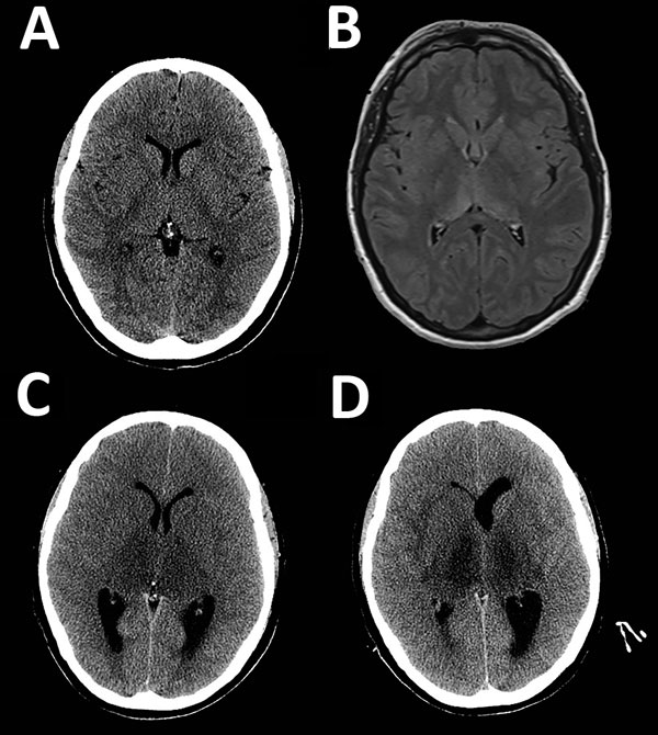
Figure 1. Neuroimaging during course of illness for a patient with a fatal infection of Murray Valley encephalitis virus imported from Australia to Canada, 2011. Each image corresponds to an axial cross-section through the thalamus and basal ganglia. A) Computed tomography (CT) at day 3. B) Magnetic resonance imaging (T2 flipped attenuation inversion recovery sequence) at day 3 showing abnormalities in the posterior thalami and splenium of the corpus callosum. C) CT when a fixed, dilated, right pupil (day 8) developed in the patient showing marked thalamic hypo-density and obstructive hydrocephalus. D) CT before death (day 10) showing necrosis of both thalami and a dilated left lateral ventricle.
Page created: January 17, 2017
Page updated: January 17, 2017
Page reviewed: January 17, 2017
The conclusions, findings, and opinions expressed by authors contributing to this journal do not necessarily reflect the official position of the U.S. Department of Health and Human Services, the Public Health Service, the Centers for Disease Control and Prevention, or the authors' affiliated institutions. Use of trade names is for identification only and does not imply endorsement by any of the groups named above.