Volume 24, Number 4—April 2018
Synopsis
Two Infants with Presumed Congenital Zika Syndrome, Brownsville, Texas, USA, 2016–2017
Figure 2

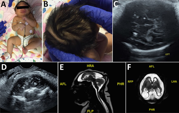
Figure 2. Term female infant (case-patient 2) with presumed congenital Zika syndrome, Brownsville, Texas, USA, 2016–2017. A) Microcephaly on the day of birth. Head circumference was 26.5 cm, which is 6.23 SDs below the mean value for term females. Craniofacial disproportion with narrow and laterally depressed frontal bone is seen. Upper wrist contractures are present, more apparent on the right, with ulnar deviation. B) Redundant scalp skin with multiple rugae. C) Transabdominal ultrasonography image of the axial transthalamic plane at 37 weeks’ gestation, showing coarse bilateral calcifications in the thalami. D) Transvaginal ultrasonography image of the coronal section at 37 weeks’ gestation, showing coarse calcifications in the thalami. E) Sagittal T2 turbo spin echo magnetic resonance image on day of birth, showing microcephaly, dysgenesis of the corpus callosum, and a small bilateral choroid plexus cyst. F) Axial T2 turbo spin echo magnetic resonance image on day of birth, showing microcephaly with enlarged extra-axial spaces and a smooth gyral pattern. Large bilateral posterior parietal and occipital lobe parenchymal cysts are present. AFL, anterior left; FLP, left posterior; HRA, right anterior; LHA, left anterior; PHR, posterior right; R, right; RFP, right posterior.
1All authors contributed equally to this article.
2Current affiliation: Texas A&M University College of Medicine, Bryan, Texas, USA.