Volume 24, Number 5—May 2018
Dispatch
Fatal Visceral Leishmaniasis Caused by Leishmania infantum, Lebanon
Figure
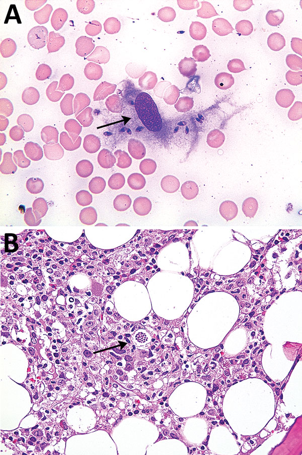
Figure. Bone marrow smear sample (A) and bone marrow aspirate (B) for patient 2 with visceral leishmaniasis caused by Leishmania infantum, Lebanon. Arrows show amastigotes within macrophages. Panel A, Wright Giemsa stain, original magnification x400; panel B, hematoxylin and eosin stain, original magnification x200.
1These senior authors contributed equally to this article.
Page created: April 17, 2018
Page updated: April 17, 2018
Page reviewed: April 17, 2018
The conclusions, findings, and opinions expressed by authors contributing to this journal do not necessarily reflect the official position of the U.S. Department of Health and Human Services, the Public Health Service, the Centers for Disease Control and Prevention, or the authors' affiliated institutions. Use of trade names is for identification only and does not imply endorsement by any of the groups named above.