Volume 25, Number 12—December 2019
Synopsis
Global Epidemiology of Buruli Ulcer, 2010–2017, and Analysis of 2014 WHO Programmatic Targets
Till F. Omansen , Alfred Erbowor-Becksen, Rie Yotsu, Tjip S. van der Werf, Alexander Tiendrebeogo, Lise Grout, and Kingsley Asiedu
Figure 1
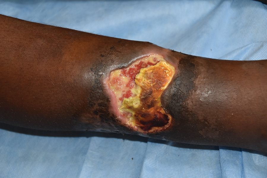
Figure 1. Typical Buruli ulcer lesion on the arm of a patient from Ghana. Central necrosis, yellowish-white slough, and undermined edges surround the wound. Photo courtesy of T.S. van der Werf.
Page created: November 18, 2019
Page updated: November 18, 2019
Page reviewed: November 18, 2019
The conclusions, findings, and opinions expressed by authors contributing to this journal do not necessarily reflect the official position of the U.S. Department of Health and Human Services, the Public Health Service, the Centers for Disease Control and Prevention, or the authors' affiliated institutions. Use of trade names is for identification only and does not imply endorsement by any of the groups named above.