Skip directly to site content Skip directly to page options Skip directly to A-Z link Skip directly to A-Z link Skip directly to A-Z link
Volume 25, Number 5—May 2019
Research

Lassa Virus Targeting of Anterior Uvea and Endothelium of Cornea and Conjunctiva in Eye of Guinea Pig Model

Joy M. Gary, Stephen R. Welch, Jana M. Ritter, JoAnn Coleman-McCray, Thanhthao Huynh, Markus H. Kainulainen, Brigid C. Bollweg, Vaunita Parihar, Stuart T. Nichol, Sherif R. Zaki, Christina F. Spiropoulou, and Jessica R. SpenglerComments to Author 
Author affiliations: Centers for Disease Control and Prevention, Atlanta, Georgia, USA

Main Article

Figure 3

Mild mononuclear anterior uveitis in eyes of guinea pigs infected with Lassa virus (LASV) Josiah by hematoxylin and eosin stain in study of LASV targeting of anterior uvea and endothelium of cornea and conjunctiva in eye. A) Anterior uvea, conjunctiva, and cornea highlighting the mild inflammation and new vessel formation in the peripheral cornea. Original magnification ×4. B) New vessel formation of the peripheral cornea. Original magnification ×12. C) New vessel formation within the cornea hig

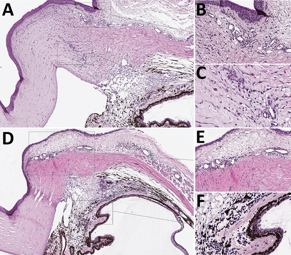
Figure 3. Mild mononuclear anterior uveitis in eyes of guinea pigs infected with Lassa virus (LASV) Josiah by hematoxylin and eosin stain in study of LASV targeting of anterior uvea and endothelium of cornea and conjunctiva in eye. A) Anterior uvea, conjunctiva, and cornea highlighting the mild inflammation and new vessel formation in the peripheral cornea. Original magnification ×4. B) New vessel formation of the peripheral cornea. Original magnification ×12. C) New vessel formation within the cornea highlighting the endothelial swelling and mixed inflammation. Original magnification ×20. D) The ciliary body, filtration angle, peripheral cornea, and a portion of the conjunctiva with mixed, mild, primarily lymphocytic inflammation in the filtration angle and around vessels in the conjunctiva, peripheral cornea, and sclera. Representative animal Jos-1. Original magnification ×6. E) Inflammation around conjunctival vessels at the margin of the cornea. Original magnification ×20. F) Mononuclear inflammation in the filtration angle and at the base of the ciliary body. Original magnification ×20. Representative animals: A–C, Jos-3; D–F, Jos-1.

Main Article

Page created: April 18, 2019
Page updated: April 18, 2019
Page reviewed: April 18, 2019
The conclusions, findings, and opinions expressed by authors contributing to this journal do not necessarily reflect the official position of the U.S. Department of Health and Human Services, the Public Health Service, the Centers for Disease Control and Prevention, or the authors' affiliated institutions. Use of trade names is for identification only and does not imply endorsement by any of the groups named above.
file_external