Skip directly to site content Skip directly to page options Skip directly to A-Z link Skip directly to A-Z link Skip directly to A-Z link
Volume 25, Number 5—May 2019
Dispatch

Novel Picornavirus in Lambs with Severe Encephalomyelitis

Leonie F. Forth, Sandra F.E. Scholes, Patricia A. Pesavento, Kenneth Jackson, Adrienne Mackintosh, Amanda Carson, Fiona Howie, Kore Schlottau, Kerstin Wernike, Anne Pohlmann, Dirk Höper, and Martin BeerComments to Author 
Author affiliations: Friedrich-Loeffler-Institut, Greifswald-Insel Riems, Germany (L.F. Forth, K. Schlottau, K. Wernike, A. Pohlmann, D. Höper, M. Beer); SRUC Veterinary Services, Edinburgh, Scotland, UK (S.F.E. Scholes, F. Howie); University of California–Davis, Davis, California, USA (P.A. Pesavento, K. Jackson); APHA Veterinary Investigation Centre, Carmarthen, Wales, UK (A. Mackintosh); APHA Veterinary Investigation Centre, Penrith, UK (A. Carson)

Main Article

Figure 1

Histologic and in situ hybridization findings in cervical segment 7 of the spinal cord of 3-week-old lamb in Scotland in study of picornavirus in lambs with severe encephalomyelitis. A) Segment view with hematoxylin and eosin stain under low power. B) Hematoxylin and eosin stain under high powerNonsuppurative myelitis is oriented on the ventral horn involving neuronal degeneration with satellitosis (black arrow), neuronophagia (arrowhead), and glial nodule formation (blue arrow), accompanied by

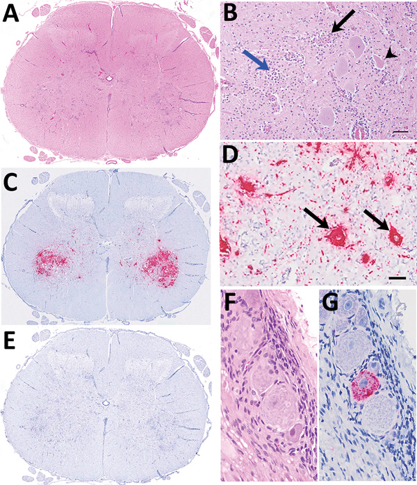
Figure 1. Histologic and in situ hybridization findings in cervical segment 7 of the spinal cord of 3-week-old lamb in Scotland in study of picornavirus in lambs with severe encephalomyelitis. A) Tranverse segmental view with hematoxylin and eosin stain under low power. B) Hematoxylin and eosin stain under high power. Nonsuppurative myelitis is oriented on the ventral horn involving neuronal degeneration with satellitosis (black arrow), neuronophagia (arrowhead), and glial nodule formation (blue arrow), accompanied by perivascular mononuclear cell accumulation and gliosis. Scale bar indicates 60 μm. C) By in situ hybridization, ovine picornavirus (OvPV) probe hybridization (red) predominates in the region of ventral horn poliomyelitis. D) Viral probe hybridization is dense in neuronal cytoplasm (black arrows) and within presumed neuronal extensions within the neuropil. Scale bar indicates 60 μm. E) Using an unrelated probe, no hybridization is detectable. F, G) Probe hybridization within scattered individual neurons within a spinal ganglion (F, hematoxylin and eosin stain; G, OvPV probe in situ hybridization). Original magnification ×400.

Main Article

Page created: April 18, 2019
Page updated: April 18, 2019
Page reviewed: April 18, 2019
The conclusions, findings, and opinions expressed by authors contributing to this journal do not necessarily reflect the official position of the U.S. Department of Health and Human Services, the Public Health Service, the Centers for Disease Control and Prevention, or the authors' affiliated institutions. Use of trade names is for identification only and does not imply endorsement by any of the groups named above.
file_external