Skip directly to site content Skip directly to page options Skip directly to A-Z link Skip directly to A-Z link Skip directly to A-Z link
Volume 25, Number 6—June 2019
Research

Novel Orthobunyavirus Causing Severe Kidney Disease in Broiler Chickens, Malaysia, 2014–2017

Vilmos PalyaComments to Author , Edit Walkóné Kovács, Szilvia Marton, Tímea Tatár-Kis, Balázs Felföldi, Barbara Forró, Marianna Domán, and Krisztián Bányai
Author affiliations: Ceva-Phylaxia Veterinary Biologicals Co. Ltd., Budapest, Hungary (V. Palya, E.W. Kovács, T. Tatár-Kis, B. Felföldi); Hungarian Academy of Sciences, Budapest (S. Marton, B. Forró, M. Domán, K. Bányai)

Main Article

Figure 7

Histologic appearance of lesions in tissues from chickens experimentally infected with a novel orthobunyavirus (Kedah fatal kidney syndrome virus) isolated from broiler chickens with severe kidney disease, Malaysia, 2014–2017. A) Kidney, showing urate deposits in the dilated Bowman’s capsule (arrows) and degeneration and dilatation of proximal convoluted tubules. Trichcrome stain; original magnification ×200. B) Liver, showing vacuolar degeneration of hepatocytes and disorganization of lobular s

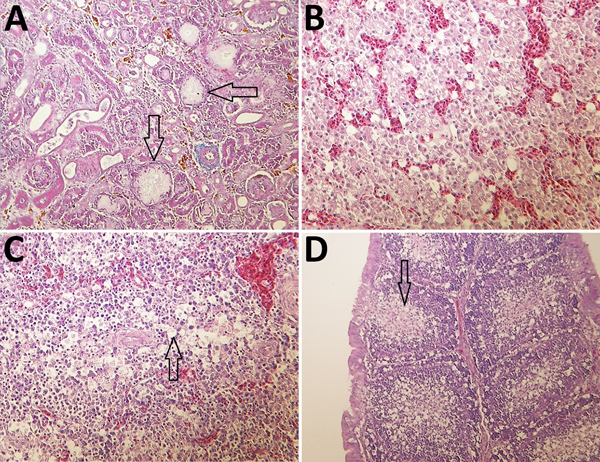
Figure 7. Histologic appearance of lesions in tissues from chickens experimentally infected with a novel orthobunyavirus (Kedah fatal kidney syndrome virus) isolated from broiler chickens with severe kidney disease, Malaysia, 2014–2017. A) Kidney, showing urate deposits in the dilated Bowman’s capsule (arrows) and degeneration and dilatation of proximal convoluted tubules. Trichcrome stain; original magnification ×200. B) Liver, showing vacuolar degeneration of hepatocytes and disorganization of lobular structure. Hematoxylin and eosin (H&E) stain; original magnification ×400. C) Spleen, showing marked lymphoid depletion and lymphocyte necrosis in the Malpighian body and vacuolation of reticulocytes in the red pulp (arrow). H&E stain; original magnification ×400. D) Bursa Fabricii, showing atrophy of bursal follicles with marked lymphoid depletion in the medullary region (arrow). H&E stain; original magnification ×200.

Main Article

Page created: May 20, 2019
Page updated: July 21, 2020
Page reviewed: July 21, 2020
The conclusions, findings, and opinions expressed by authors contributing to this journal do not necessarily reflect the official position of the U.S. Department of Health and Human Services, the Public Health Service, the Centers for Disease Control and Prevention, or the authors' affiliated institutions. Use of trade names is for identification only and does not imply endorsement by any of the groups named above.
file_external