Skip directly to site content Skip directly to page options Skip directly to A-Z link Skip directly to A-Z link Skip directly to A-Z link
Volume 25, Number 8—August 2019
Dispatch

Kaposi Sarcoma in Mantled Guereza

Anna GrewerComments to Author , Martina Bleyer, Kerstin Mätz-Rensing, Alexander S. Hahn, Tim Rüggeberg, Gregor Babaryka, Andre Zimmermann, Stefan Pöhlmann, and Artur Kaul
Author affiliations: Zoo Krefeld GmbH, Krefeld, Germany (A. Grewer); German Primate Center–Leibniz Institute for Primate Research, Göttingen, Germany (M. Bleyer, K. Mätz-Rensing, A.S. Hahn, S. Pöhlmann, A. Kaul); Heinrich-Heine-University Düsseldorf, Düsseldorf, Germany (T. Rüggeberg, G. Babaryka, A. Zimmermann); Joint Practice for Oral and Maxillofacial Surgery, Neuss, Germany (A. Zimmermann); University of Göttingen, Göttingen (S. Pöhlmann)

Main Article

Figure 1

Disease manifestations in mantled guereza with Kaposi sarcoma. A) Oligofocal flattened masses on the inner aspects of the lower lip. B) Multinodular fissured masses at the gingival margin. C) Fibrovascular stroma in the subepithelial propria of the lower lip with spindle cell proliferations delineating narrow vascular clefts and containing lymphoplasmacytic inflammatory cell infiltrates, hematoxylin and eosin stained; scale bar indicates 200 µm. D) Spindle cell proliferation with cavern formatio

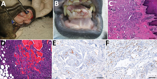
Figure 1. Disease manifestations in mantled guereza with Kaposi sarcoma. A) Oligofocal flattened masses on the inner aspects of the lower lip. B) Multinodular fissured masses at the gingival margin. C) Fibrovascular stroma in the subepithelial propria of the lower lip with spindle cell proliferations delineating narrow vascular clefts and containing lymphoplasmacytic inflammatory cell infiltrates, hematoxylin and eosin stained; scale bar indicates 200 µm. D) Spindle cell proliferation with cavern formation in the perinodal adipose tissue of the mandibular lymph node; hematoxylin and eosin stained; scale bar indicates 100 µm. E) Immunohistochemical staining showing variable Ki67 expression in <20% of spindle cells, streptavidin-biotin complex method–diaminobenzidine tetrahydrochloride; scale bar indicates 100 µm. F) Immunohistochemical staining showing nuclear expression of latent nuclear antigen 1 in ≈50%–60% of spindle cells, streptavidin-biotin complex method–diaminobenzidine tetrahydrochloride; scale bar indicates 50 µm.

Main Article

Page created: July 16, 2019
Page updated: July 16, 2019
Page reviewed: July 16, 2019
The conclusions, findings, and opinions expressed by authors contributing to this journal do not necessarily reflect the official position of the U.S. Department of Health and Human Services, the Public Health Service, the Centers for Disease Control and Prevention, or the authors' affiliated institutions. Use of trade names is for identification only and does not imply endorsement by any of the groups named above.
file_external