Volume 26, Number 8—August 2020
Dispatch
Plasma-Derived Extracellular Vesicles as Potential Biomarkers in Heart Transplant Patient with Chronic Chagas Disease
Nuria Cortes-Serra, Maria Tays Mendes, Clara Mazagatos1, Joan Segui-Barber, Cameron C. Ellis, Cristina Ballart, Ana Garcia-Alvarez, Montserrat Gállego, Joaquim Gascon, Igor C. Almeida, María Jesús Pinazo , and Carmen Fernandez-Becerra
Figure 1
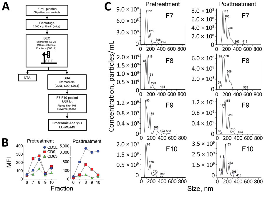
Figure 1. Isolation and characterization of plasma-derived EVs. A) Schematic diagram of the isolation and characterization of EVs derived from plasma samples. The details of each step are explained in The Study section. B) EVs were characterized by BBA using the classical EV markers CD5L, CD9, and CD63. C) NTA of SEC fractions F7–10. BBA, bead-based assay; EV, extracellular vesicle; LC-MS/MS, liquid chromatography–tandem mass spectrometry; MFI, median fluorescence intensity; NTA, nanoparticle tracking analysis; SEC, size-exclusion chromatography.
1Current affiliation: CIBER Epidemiología y Salud Pública (CIBERESP); Centro Nacional de Epidemiología, Instituto de Salud Carlos III, Madrid, Spain.
Page created: April 27, 2020
Page updated: July 18, 2020
Page reviewed: July 18, 2020
The conclusions, findings, and opinions expressed by authors contributing to this journal do not necessarily reflect the official position of the U.S. Department of Health and Human Services, the Public Health Service, the Centers for Disease Control and Prevention, or the authors' affiliated institutions. Use of trade names is for identification only and does not imply endorsement by any of the groups named above.