Skip directly to site content Skip directly to page options Skip directly to A-Z link Skip directly to A-Z link Skip directly to A-Z link
Volume 27, Number 1—January 2021
Dispatch

In Vivo Observation of Cutaneous Larva Migrans by Fluorescence-Advanced Videodermatoscopy

Alice Ramondetta1Comments to Author , Simone Ribero1, Pietro Quaglino2, and Paolo Broganelli2
Author affiliation: Università degli Studi di Torino, Turin, Italy

Main Article

Figure

Imaging and biopsy results for patient with cutaneous larva migrans, Turin, Italy. A) Fluorescence-advanced videodermatoscopy showed larva with a diameter of 70–80 μm, located intraepidermally, ≈0.5 cm to the right of the distal end of the serpentine path (indicated in the inset) caused by its passage in the skin. White arrow indicates head of the larva (original magnification ×500). B) Hematoxylin and eosin–stained longitudinal skin section obtained from a 4-mm biopsy specimen, showing the cavity at the epidermal level in which it is possible to observe the larva inside (original magnification ×20). Once the biopsy specimen was obtained, confirmation of a larva by morphologic details or molecular techniques was necessary to differentiate animal nematodes from other larvae, particularly Strongyloides stercoralis, because treatment and follow-up would be different.

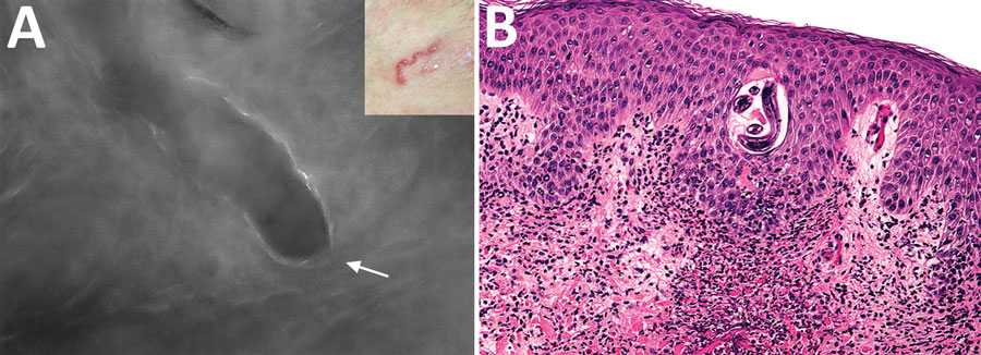
Figure. Imaging and biopsy results for patient with cutaneous larva migrans, Turin, Italy. A) Fluorescence-advanced videodermatoscopy showed larva with a diameter of 70–80 μm, located intraepidermally, ≈0.5 cm to the right of the distal end of the serpentine path (indicated in the inset) caused by its passage in the skin. White arrow indicates head of the larva (original magnification ×500). B) Hematoxylin and eosin–stained longitudinal skin section obtained from a 4-mm biopsy specimen, showing the cavity at the epidermal level in which it is possible to observe the larva inside (original magnification ×20). Once the biopsy specimen was obtained, confirmation of a larva by morphologic details or molecular techniques was necessary to differentiate animal nematodes from other larvae, particularly Strongyloides stercoralis, because treatment and follow-up would be different.

Main Article

1These authors contributed equally to this article.

2These senior authors contributed equally to this article.

Page created: September 15, 2020
Page updated: December 21, 2020
Page reviewed: December 21, 2020
The conclusions, findings, and opinions expressed by authors contributing to this journal do not necessarily reflect the official position of the U.S. Department of Health and Human Services, the Public Health Service, the Centers for Disease Control and Prevention, or the authors' affiliated institutions. Use of trade names is for identification only and does not imply endorsement by any of the groups named above.
file_external