Skip directly to site content Skip directly to page options Skip directly to A-Z link Skip directly to A-Z link Skip directly to A-Z link
Volume 27, Number 3—March 2021
Dispatch

Lung Pathology of Mutually Exclusive Co-infection with SARS-CoV-2 and Streptococcus pneumoniae

Tetsuya Tsukamoto1, Noriko Nakajima1, Aki Sakurai1, Masayuki Nakajima, Eiko Sakurai, Yuko Sato, Kenta Takahashi, Takayuki Kanno, Michiko Kataoka, Harutaka Katano, Mitsunaga Iwata, Yohei Doi, and Tadaki SuzukiComments to Author 
Author affiliations: Fujita Health University School of Medicine, Toyoake, Japan (T. Tsukamoto, A. Sakurai, M. Nakajima, E. Sakurai, M. Iwata, Y. Doi); National Institute of Infectious Diseases, Tokyo, Japan (N. Nakajima, Y. Sato, K. Takahashi, T. Kanno, M. Kataoka, H. Katano, T. Suzuki)

Main Article

Figure 2

Microscopic findings of the lungs of a patient in Japan co-infected with SARS-CoV-2 and Streptococcus pneumoniae. A) Histopathology of lung section R12 (shown in Figure 1). Scale bar indicates 2 mm. B) Magnified image of the black square (top left) in panel A: exudative phase of diffuse alveolar damage (DAD) with hyaline membranes. Scale bar indicates 100 μm. C) Magnified image of the red square (bottom right) in panel A: edema and bronchopneumonia with massive infiltration of neutrophils in the alveolar spaces. Scale bar indicates 100 μm. D, E) Magnified images of the same areas of consecutive sections as B and C, respectively, showing SARS-CoV-2 antigen stained green (Vina green) and S. pneumoniae antigen stained brown (3,3′-diaminobenzidine) by enzyme-labeled double immunohistochemistry. The SARS-CoV-2 antigens were detected predominantly in the DAD area (D; scale bar indicates 50 μm). The S. pneumoniae antigens were detected predominantly in the bronchopneumonia area (E; scale bar indicates 50 μm). Insets show magnified images of the staining cells (scale bars indicate 10 μm).

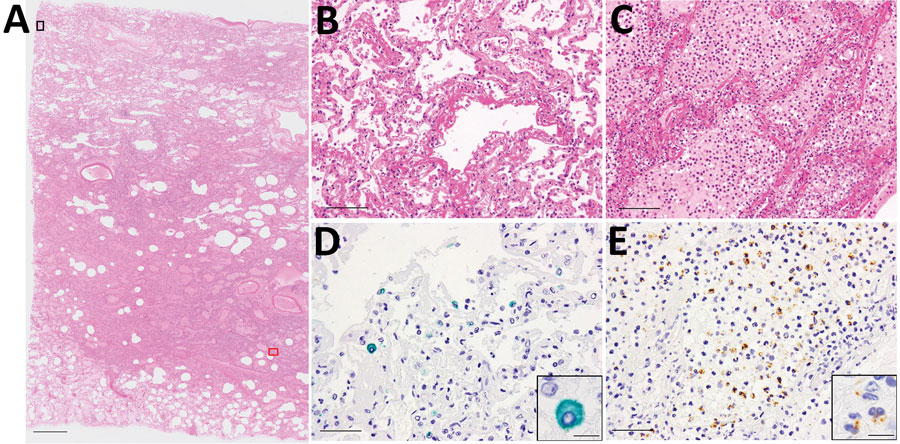
Figure 2. Microscopic findings of the lungs of a patient in Japan co-infected with SARS-CoV-2 and Streptococcus pneumoniae. A) Histopathology of lung section R12 (shown in Figure 1). Scale bar indicates 2 mm. B) Magnified image of the black square (top left) in panel A: exudative phase of diffuse alveolar damage (DAD) with hyaline membranes. Scale bar indicates 100 μm. C) Magnified image of the red square (bottom right) in panel A: edema and bronchopneumonia with massive infiltration of neutrophils in the alveolar spaces. Scale bar indicates 100 μm. D, E) Magnified images of the same areas of consecutive sections as B and C, respectively, showing SARS-CoV-2 antigen stained green (Vina green) and S. pneumoniae antigen stained brown (3,3′-diaminobenzidine) by enzyme-labeled double immunohistochemistry. The SARS-CoV-2 antigens were detected predominantly in the DAD area (D; scale bar indicates 50 μm). The S. pneumoniae antigens were detected predominantly in the bronchopneumonia area (E; scale bar indicates 50 μm). Insets show magnified images of the staining cells (scale bars indicate 10 μm).

Main Article

1These authors contributed equally to this article.

Page created: January 06, 2021
Page updated: February 21, 2021
Page reviewed: February 21, 2021
The conclusions, findings, and opinions expressed by authors contributing to this journal do not necessarily reflect the official position of the U.S. Department of Health and Human Services, the Public Health Service, the Centers for Disease Control and Prevention, or the authors' affiliated institutions. Use of trade names is for identification only and does not imply endorsement by any of the groups named above.
file_external