Volume 27, Number 4—April 2021
Research Letter
Co-infection with Severe Fever with Thrombocytopenia Syndrome Virus and Rickettsia japonica after Tick Bite, Japan
Abstract
Severe fever with thrombocytopenia syndrome was diagnosed in a febrile woman in Japan after a tick bite. However, Rickettsia japonica DNA was retrospectively detected in the eschar specimen, suggesting co-infection from the bite. Establishment of the severe fever with thrombocytopenia syndrome virus infection might have overpowered the R. japonica infection.
Severe fever with thrombocytopenia syndrome (SFTS) is caused by SFTS virus (SFTSV), a novel phlebovirus in the family Bunyaviridae (1). It has been reported that SFTS is endemic to Japan (2). SFTS is classified as a viral hemorrhagic fever, and its case-fatality rate in Japan is ≈30% (3).
Japanese spotted fever (JSF) is an acute tickborne rickettsiosis caused by Rickettsia japonica and is endemic to Japan (4). Most cases of SFTS in Japan have been reported in southwestern Japan, and the JSF-endemic area overlaps the areas to which SFTS is endemic. Because the Haemaphysalis longicornis tick is a vector for both SFTSV and R. japonica (4,5), co-infection events might occur in patients with SFTS or R. japonica infection.
Figure
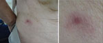
Figure. Eschar at site of tick bite surrounded by exanthema on lower right back of patient with severe fever with thrombocytopenia syndrome and positive Rickettsia japonica DNA in eschar, Japan. ...
A woman 84 years of age was bitten on her lower right back by a tick while working in a field. She became febrile on day 1, experienced mild delirium on day 2, and visited the emergency department of Mitoyo General Hospital (Kanonji, Japan) on day 5, where she had low-grade fever but was alert and lucid. Physical examination revealed an eschar surrounded by exanthema on her lower right back (Figure). She had noticed the eschar on the day after the bite, and her family removed it. We observed no other skin exanthema on her body. Laboratory analysis revealed thrombocytopenia and leukocytopenia (Table). Serum chemistry analyses revealed elongation of the activated partial thromboplastin time and an increase in the D-dimer level, suggesting coagulopathy. Because increases in aspartate transaminase and blood urea nitrogen were noted, liver and renal functions might have been impaired transiently (Table).
Because of the fever, thrombocytopenia, history of tick bite, and eschar with localized exanthema, we suspected JSF. The patient’s blood samples and the crust of the eschar were tested by PCR assays for R. japonica, Orientia tsutsugamushi, and SFTSV. The serum sample tested positive for SFTSV by a conventional 1-step reverse transcription PCR reported previously (6). R. japonica DNA was also detected in the eschar sample through the methods described previously (7), but it was not detected in serum samples. We empirically administered 100 mg of minocycline intravenously for 7 days, after which minocycline was administered orally every 12 hours for 3 days. Her symptoms resolved without complications by day 6, the second day of admission. After discharge from the hospital on day 12, outpatient follow-up was uneventful.
We analyzed blood specimens to examine paired serum antibody titers against SFTSV in the acute phase and convalescent phase with indirect immunofluorescence assay (IFA) (8), which indicated a substantial increase in the antibody to SFTSV from <10 to 640. A relatively low level of viremia (154 copies/mL) was also confirmed in the acute phase (day 4) of the disease by quantitative PCR assays (6). We tested paired serum from the acute phase (day 4) and the convalescent phase (day 27) for IgG and IgM titers against R. japonica by IFA as described previously (9). IgG and IgM against R. japonica were not detected in either the acute-phase or convalescent-phase serum samples. This result suggests that a general R. japonica infection had not established itself and that infection was localized to the eschar, in which erythematous lesions were present, and R. japonica DNA was detected only in the eschar sample (Figure). Unfortunately, the nucleotide sequence of the R. japonica genome amplified from the eschar was not determined.
The clinical course and laboratory results of this patient, with the exception of the eschar, were consistent with SFTS but not JSF. It has been reported that a tick bite scar could not be found in 56% of SFTS patients (6), whereas skin eruptions appear in 100% of patients with JSF and tick bite eschar appear in 90% of patients with JSF (10). The patient showed no other skin eruptions besides the eschar at the site of the tick bite (Figure). It is highly possible that the eschar on this patient could have been caused by an inflammatory response induced by the local R. japonica infection. R. japonica did not induce systemic symptoms in this patient for 2 possible reasons. First, the incubation time for SFTS might be shorter than that of JSF. Second, the initiation of antimicrobial drugs in the early phase of disease might have ameliorated the clinical course of the diseases.
In conclusion, we describe a patient with a generalized SFTSV infection and a localized skin lesion caused by R. japonica at the site of a tick bite. This study suggests that SFTS patients with eschar at the site of a tick bite should be treated with appropriate antimicrobial drugs, such as doxycycline and minocycline.
Dr. Fujikawa is a chief director in the Department of General Internal Medicine, Kagawa, Mitoyo General Hospital, Japan. His research interests include general internal medicine and medical education.
Acknowledgments
The study was carried out partially with financial support from the Japan Agency for Medical Research and Development (AMED, 19fk0108081j and 20fk0108081j). The funders had no role in study design, data collection and analysis, decision to publish, or preparation of the manuscript.
T.F. and K.Y. contributed to clinical management and writing of the manuscript. M.S. contributed to writing of the manuscript. T.Y., T.K., and M.S. contributed to the virological diagnosis. All authors had full access to all data in the study and all take responsibility for the integrity of the data and the accuracy of the data analysis.
References
- Yu XJ, Liang MF, Zhang SY, Liu Y, Li JD, Sun YL, et al. Fever with thrombocytopenia associated with a novel bunyavirus in China. N Engl J Med. 2011;364:1523–32. DOIPubMedGoogle Scholar
- Takahashi T, Maeda K, Suzuki T, Ishido A, Shigeoka T, Tominaga T, et al. The first identification and retrospective study of Severe Fever with Thrombocytopenia Syndrome in Japan. J Infect Dis. 2014;209:816–27. DOIPubMedGoogle Scholar
- Kato H, Yamagishi T, Shimada T, Matsui T, Shimojima M, Saijo M, et al.; SFTS epidemiological research group-Japan. SFTS epidemiological research group-Japan. Epidemiological and clinical features of severe fever with thrombocytopenia syndrome in Japan, 2013–2014. PLoS One. 2016;11:
e0165207 . DOIPubMedGoogle Scholar - Yamaji K, Aonuma H, Kanuka H. Distribution of tick-borne diseases in Japan: Past patterns and implications for the future. J Infect Chemother. 2018;24:499–504. DOIPubMedGoogle Scholar
- Saijo M. Circulation of severe fever with thrombocytopenia syndrome virus (SFTSV) in nature: transmission of SFTSV between mammals and ticks. In: Saijo M, editor. Severe fever with thrombocytopenia syndrome. Singapore: Springer Nature Singapore Pte Ltd.; 2019. p. 151–72.
- Yoshikawa T, Fukushi S, Tani H, Fukuma A, Taniguchi S, Toda S, et al. Sensitive and specific PCR systems for detection of both Chinese and Japanese severe fever with thrombocytopenia syndrome virus strains and prediction of patient survival based on viral load. J Clin Microbiol. 2014;52:3325–33. DOIPubMedGoogle Scholar
- Furuya Y, Katayama T, Yoshida Y, Kaiho I. Specific amplification of Rickettsia japonica DNA from clinical specimens by PCR. J Clin Microbiol. 1995;33:487–9. DOIPubMedGoogle Scholar
- Fukuma A, Fukushi S, Yoshikawa T, Tani H, Taniguchi S, Kurosu T, et al. Severe fever with thrombocytopenia syndrome virus antigen detection using monoclonal antibodies to the nucleocapsid protein. PLoS Negl Trop Dis. 2016;10:
e0004595 . DOIPubMedGoogle Scholar - Uchiyama T, Zhao L, Yan Y, Uchida T. Cross-reactivity of Rickettsia japonica and Rickettsia typhi demonstrated by immunofluorescence and Western immunoblotting. Microbiol Immunol. 1995;39:951–7. DOIPubMedGoogle Scholar
- Mahara F. Japanese spotted fever: report of 31 cases and review of the literature. Emerg Infect Dis. 1997;3:105–11. DOIPubMedGoogle Scholar
Figure
Table
Cite This ArticleOriginal Publication Date: March 10, 2021
Table of Contents – Volume 27, Number 4—April 2021
| EID Search Options |
|---|
|
|
|
|
|
|
Please use the form below to submit correspondence to the authors or contact them at the following address:
Tatsuya Fujikawa, Department of General Internal Medicine, Mitoyo General Hospital, 708 Himehama Toyohama, Kanonji, Kagawa 769-1695, Japan
Top