Skip directly to site content Skip directly to page options Skip directly to A-Z link Skip directly to A-Z link Skip directly to A-Z link
Volume 27, Number 5—May 2021
Research Letter

Intersecting Paths of Emerging and Reemerging Infectious Diseases

Tais M. Wilson, Christopher D. Paddock, Sarah Reagan-Steiner, Julu Bhatnagar, Roosecelis B. Martines, Andrea L. Wiens, Michael Madsen, Kenneth K. Komatsu, Heather Venkat, and Sherif R. ZakiComments to Author 
Author affiliations: Universidade de Brasília, Brasília, Brazil (T.M. Wilson); Centers for Disease Control and Prevention, Atlanta, Georgia, USA (T.M. Wilson, C.D. Paddock, S. Reagan-Steiner, J. Bhatnagar, R.B. Martines, H. Venkat, S.R. Zaki); Pinal County Office of the Medical Examiner, Florence, Arizona, USA (A.L. Wiens); Coconino County Health and Human Services Medical Examiner’s Office, Flagstaff, Arizona, USA (M. Madsen); Arizona Department of Health Services, Phoenix, Arizona, USA (K.K. Komatsu, H. Venkat)

Main Article

Figure

Histopathologic and immunohistochemical characteristics of fatal hantavirus pulmonary syndrome in 2 patients, Arizona, USA, 2020. A) Patient 1 lung tissue, showing intravascular leukocytosis with left shift (left, original magnification ×50) and hantavirus antigen immunostaining (red) in pulmonary microvasculature (right, original magnification ×158). B) Patient 1 spleen tissue, showing immunoblast proliferation in the red pulp and periarteriolar sheaths (left, original magnification ×50) and immunoblasts with high nuclear to cytoplasmic ratio, vesicular and prominent nucleoli (arrows) and mitosis (arrowhead) (right, original magnification ×158). C) Patient 2 lung tissue, showing severe intraalveolar edema (original magnification ×12.5). D) Patient 2 lung tissue, showing interstitial pneumonitis with hyaline membranes (arrows) (original magnification ×50). E) Patient 2 lung tissue, showing hantavirus antigen immunostaining (red) in pulmonary microvasculature (left, original magnification ×50; right, original magnification ×158). F) Patient 2 kidney tissue, showing hantavirus antigen immunostaining (red) in glomerular capillaries (arrowhead) and interstitial vessel (arrow) (original magnification ×100).

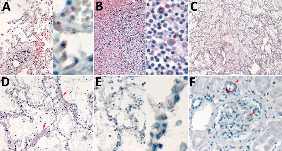
Figure. Histopathologic and immunohistochemical characteristics of fatal hantavirus pulmonary syndrome in 2 patients, Arizona, USA, 2020. A) Patient 1 lung tissue, showing intravascular leukocytosis with left shift (left, original magnification ×50) and hantavirus antigen immunostaining (red) in pulmonary microvasculature (right, original magnification ×158). B) Patient 1 spleen tissue, showing immunoblast proliferation in the red pulp and periarteriolar sheaths (left, original magnification ×50) and immunoblasts with high nuclear to cytoplasmic ratio, vesicular and prominent nucleoli (arrows) and mitosis (arrowhead) (right, original magnification ×158). C) Patient 2 lung tissue, showing severe intraalveolar edema (original magnification ×12.5). D) Patient 2 lung tissue, showing interstitial pneumonitis with hyaline membranes (arrows) (original magnification ×50). E) Patient 2 lung tissue, showing hantavirus antigen immunostaining (red) in pulmonary microvasculature (left, original magnification ×50; right, original magnification ×158). F) Patient 2 kidney tissue, showing hantavirus antigen immunostaining (red) in glomerular capillaries (arrowhead) and interstitial vessel (arrow) (original magnification ×100).

Main Article

Page created: March 06, 2021
Page updated: April 22, 2021
Page reviewed: April 22, 2021
The conclusions, findings, and opinions expressed by authors contributing to this journal do not necessarily reflect the official position of the U.S. Department of Health and Human Services, the Public Health Service, the Centers for Disease Control and Prevention, or the authors' affiliated institutions. Use of trade names is for identification only and does not imply endorsement by any of the groups named above.
file_external