Volume 29, Number 5—May 2023
Dispatch
Poor Prognosis for Puumala Virus Infections Predicted by Lymphopenia and Dyspnea
Abstract
We investigated a prospective cohort of 23 patients who had Puumala virus infection in Austria to determine predictors of infection outcomes. We reviewed routinely available clinical and laboratory parameters collected when patients initially sought care. Low absolute lymphocyte count and dyspnea were parameters associated with a severe course of infection.
Hantaviruses are emerging rodentborne pathogens that cause clinical illness in humans. During the past few decades, hantavirus infection outbreaks increased, demonstrating an emerging problem for healthcare systems (1). Human hantavirus infections cause 2 well-defined clinical syndromes: hemorrhagic fever with renal syndrome, caused by Old World hantaviruses originating in Europe and Asia; and hantavirus cardiopulmonary syndrome. Hemorrhagic fever with renal syndrome is characterized by acute renal failure, thrombocytopenia, and mortality rates of 0.1%‒0.4%, and hantavirus cardiopulmonary syndrome is characterized by severe involvement of the respiratory and circulatory systems and case-fatality rates >30% (1).
Most cases of infection with hantavirus in Europe are caused by Puumala virus (PUUV) (2,3). Clinical manifestations of PUUV infection vary from subclinical, mild and moderate to severe, with an urgent need for intensive care treatment. However, biomarkers or clinical parameters for risk stratification of PUUV infection are lacking. In this prospective cohort study, we aimed to clarify the prognostic value of routinely assessable clinical and laboratory values in PUUV infection requiring hospital admission.
This study was approved by the institutional review board of the Medical University of Graz (approval no. 33-329 ex 20/21). Written informed consent was obtained from all participants.
We performed a prospective, observational, pilot study, enrolling all consecutive adult patients admitted to the Department of Internal Medicine, Medical University of Graz, Austria, because of clinical suspicion of PUUV infection and detection of PUUV IgM by using point-of-care testing (Reagena POC PUUMALA IgM, https://www.reagena.com). We used a PUUV reverse transcription PCR as a confirmation test as described (2). Patient data were uniformly collected as described (4). We obtained laboratory, clinical, and radiologic data from our in-house electronic healthcare database system and from handwritten charts and inserted these data into a predefined electronic case report form by using REDCap electronic data capture (5,6). We defined a severe course of PUUV infection if a patient needed oxygen (<92% blood oxygen saturation while breathing ambient air) or hemodialysis or intensive care unit admission.
We performed statistical analyses by using Stata version 16.1 (StataCorp., https://www.stata.com). We report continuous data as medians (25th–75th percentiles) and summarized categorical data by using absolute frequencies and percentages. We applied rank-sum tests, χ2 tests, and Fisher exact tests to investigate the association between 1 continuous variable and 1 categorical variable and between 2 categorical variables. To identify variables associated with a severe course of PUUV infection among the 25 variables that were collected in the presence of multiple testing, we prespecified a Sidák corrected α of association, resulting in p values <0.002 to indicate statistical significance. We used logistic models for univariate and multivariable modeling of PUUV infection severity and assessed the optimal cutoff to separate patients with and without severe course of PUUV infection by using a maximized Youdens index within a receiver operating characteristic analysis. The follow-up time was truncated at 90 days after diagnosis because no events were expected after this time interval. We computed overall survival of the cohort by using Kaplan-Meier estimators.
A total of 23 patients were included in the analysis (Table). Median age at diagnosis was 49 years (25th‒75th percentile 34–59 years), and 6 (23%) patients were women. Five (22%) patients had underlying conditions (Table). Ten (44%) patients reported activities with a predisposition to rodents or rodent excreta. The most frequent symptom at diagnosis was fever, which was observed in 100% of the patients. The median body temperature at diagnosis was 39.5°C (25th–75th percentiles 39.0°C–39.7°C). Most (17/23, 74%) patients had headache and reported concomitant use of analgesics. All 3 patients who had dyspnea came to the hospital because of this primary symptom. Symptom duration was 2 (25th‒75th percentile 1–3) days before admission to the hospital.
A severe course of the PUUV infection was observed in 5 (22%) of 23 patients. For these 5 patients, severe PUUV infection was diagnosed by need for oxygen therapy (n = 5), intensive care unit admission (n = 4), and renal replacement therapy (n = 2). Two of those 5 patients had to be treated with extracorporeal membrane oxygenation. Median length of in-patient hospital stay was 7 (25th–75th percentiles 4–12) days. One patient died from multiorgan failure after PUUV infection, corresponding to a 90-day overall survival rate of 96% (95% CI 73%–99%).
Figure
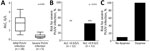
Figure. Poor prognosis for PUUV infections predicted by lymphopenia and dyspnea. A) Box plot showing difference in ALC between patients who had a mild clinical course and those who had a...
Dyspnea, predisposition to contact with rodents, shortened prothrombin time, and low absolute lymphocyte (ALC) count when patients sought care were associated with severe PUUV infection at the 5% level. However, because of the prespecified α corrected for multiple testing, we identified only dyspnea and a low ALC as major predictors for this outcome. Median ALC levels were 1.8 giga (G)/L (25th–75th percentiles 0.9–2.4 G/L) for patients who had nonsevere PUUV and 0.3 G/L (25th–75th percentiles 0.2–0.5 G/L) for patients who had severe PUUV (p<0.0001) (Figure, panel A).
Univariable logistic regression showed that a 0.1 G/L decrease in ALC predicted a 2.3-fold increase in risk for severe PUUV infection (odds ratio 2.28, 95% CI 1.03–5.03; p = 0.042). The area under the receiver operating characteristic curve for ALC for discriminating between patients with and without severe PUUV infection was 0.97, and an ALC cutoff <0.9 G/L was computed for identifying patients at high risk for severe PUUV infection. The risk for severe PUUV infection was 0% for the 12 patients who had an ALC >0.9 G/L and 45% for the 11 patients who had an ALC <0.9 G/L (p = 0.014) (Figure, panel B).
For the 20 patients without dyspnea at initial evaluation, 2 (10%) cases of severe PUUV infection were observed. In contrast, 3/3 (100%) patients with dyspnea at baseline showed development of severe PUUV infection (p = 0.006) (Figure, panel C).
Risk stratification is crucial for personalized medicine and optimized patient allocation, especially if outpatient management might be considered. Because PUUV has a benign course in most cases, easily assessable predictors of the disease course support clinicians in their decision on patient management (7–9). We report a small but well-characterized prospective patient cohort, demonstrating lymphopenia and dyspnea at time of first medical contact and diagnosis of PUUV infection as predictors of adverse clinical course.
Most studies focusing on risk stratification in PUUV infection reported kidney injury and renal failure as clinical endpoints, although PUUV infection causes a much wider syndrome, including renal failure, respiratory failure, bleeding events, and circulatory failure (8,10,11). Therefore, we analyzed a composite endpoint consisting of all relevant complications of PUUV infection to support clinical decisions for admission or outpatient care.
A recent review summarized severity biomarkers in PUUV orthohantavirus infection (12). However, almost all mentioned biomarkers are difficult to measure, need special laboratory platforms, or are cost-intensive. In our study, we observed that low ALC and dyspnea are easily accessible markers of poor outcomes in an unbiased approach. In our approach, we used all laboratory parameters, signs, and symptoms observed at first medical contact and meticulously corrected for multiplicity. However, the case number of our study was limited, and the parameters should be seen as clinical warning signs of a potential severe clinical course of PUUV infection. The proposed biomarkers need further validation in independent cohorts to prove their utility in a clinical setting.
Our investigated biomarkers were limited to PUUV-infected patients admitted to the hospital, as described in our inclusion strategy. Because only hospitalized patients were included, the observed mortality rate of 4% is slightly higher than rates reported in epidemiologic studies (1,13).
In summary, we report a prospective cohort of 23 patients who had PUUV infection in an endemic area in central Europe. Our findings indicate that low ALC and dyspnea are parameters associated with a severe course of PUUV infection.
Dr. Hatzl is a hematology and intensive care consultant at the Department of Internal Medicine, Medical University of Graz, Graz, Austria. His primary research interest is emerging viral diseases and infectious diseases in critically ill and immunocompromised patients.
S.H., F.K., and R.K. designed the study; S.H. and M.L. collected clinical data; S.H. performed statistical analysis; and S.H. and R.K. analyzed results and wrote the first draft of the manuscript. All authors reviewed the draft and approved the final version.
Acknowledgment
The study was supported by the Styrian government (project no. ABT12-106729/2022-13).
References
- Avšič-Županc T, Saksida A, Korva M. Hantavirus infections. Clin Microbiol Infect. 2019;21S:e6–16. DOIPubMedGoogle Scholar
- Camp JV, Schmon E, Krause R, Sixl W, Schmid D, Aberle SW. Genetic diversity of Puumala orthohantavirus in rodents and human patients in Austria, 2012–2019. Viruses. 2021;13:640. DOIPubMedGoogle Scholar
- Heyman P, Ceianu CS, Christova I, Tordo N, Beersma M, João Alves M, et al. A five-year perspective on the situation of haemorrhagic fever with renal syndrome and status of the hantavirus reservoirs in Europe, 2005-2010. Euro Surveill. 2011;16:19961. DOIPubMedGoogle Scholar
- Hatzl S, Reisinger AC, Posch F, Prattes J, Stradner M, Pilz S, et al. Antifungal prophylaxis for prevention of COVID-19-associated pulmonary aspergillosis in critically ill patients: an observational study. Crit Care. 2021;25:335. DOIPubMedGoogle Scholar
- Harris PA, Taylor R, Thielke R, Payne J, Gonzalez N, Conde JG. Research electronic data capture (REDCap)—a metadata-driven methodology and workflow process for providing translational research informatics support. J Biomed Inform. 2009;42:377–81. DOIPubMedGoogle Scholar
- Harris PA, Taylor R, Minor BL, Elliott V, Fernandez M, O’Neal L, et al.; REDCap Consortium. The REDCap consortium: Building an international community of software platform partners. J Biomed Inform. 2019;95:
103208 . DOIPubMedGoogle Scholar - Vaheri A, Strandin T, Hepojoki J, Sironen T, Henttonen H, Mäkelä S, et al. Uncovering the mysteries of hantavirus infections. Nat Rev Microbiol. 2013;11:539–50. DOIPubMedGoogle Scholar
- Vaheri A, Henttonen H, Mustonen J. Hantavirus research in Finland: highlights and perspectives. Viruses. 2021;13:1452. DOIPubMedGoogle Scholar
- Hjertqvist M, Klein SL, Ahlm C, Klingstrom J. Mortality rate patterns for hemorrhagic fever with renal syndrome caused by Puumala virus. Emerg Infect Dis. 2010;16:1584–6. DOIPubMedGoogle Scholar
- Mustonen J, Mäkelä S, Outinen T, Laine O, Jylhävä J, Arstila PT, et al. The pathogenesis of nephropathia epidemica: new knowledge and unanswered questions. Antiviral Res. 2013;100:589–604. DOIPubMedGoogle Scholar
- Mustonen J, Outinen T, Laine O, Pörsti I, Vaheri A, Mäkelä S. Kidney disease in Puumala hantavirus infection. Infect Dis (Lond). 2017;49:321–32. DOIPubMedGoogle Scholar
- Outinen TK, Mäkelä S, Pörsti I, Vaheri A, Mustonen J. Severity biomarkers in Puumala hantavirus infection. Viruses. 2021;14:45. DOIPubMedGoogle Scholar
- Mittler E, Wec AZ, Tynell J, Guardado-Calvo P, Wigren-Byström J, Polanco LC, et al. Human antibody recognizing a quaternary epitope in the Puumala virus glycoprotein provides broad protection against orthohantaviruses. Sci Transl Med. 2022;14:
eabl5399 . DOIPubMedGoogle Scholar
Figure
Table
Cite This ArticleOriginal Publication Date: April 13, 2023
Table of Contents – Volume 29, Number 5—May 2023
| EID Search Options |
|---|
|
|
|
|
|
|
Please use the form below to submit correspondence to the authors or contact them at the following address:
Robert Krause, Division of Infectious Diseases, Department of Internal Medicine, Medical University of Graz, Auenbruggerplatz 15, Graz, Austria
Top