Volume 30, Number 3—March 2024
Research
Microsporidia (Encephalitozoon cuniculi) in Patients with Degenerative Hip and Knee Disease, Czech Republic
Bohumil Sak , Petra Gottliebová, Elka Nyčová, Nikola Holubová, Jana Fenclová, Marta Kicia, Żaneta Zajączkowska, and Martin Kváč
Figure 3
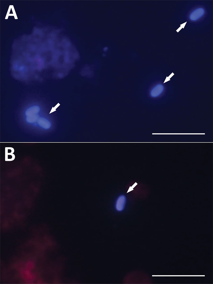
Figure 3. Microscopic analysis of Encephalitozoon cuniculi spores isolated from immunocompetent patients in study of microsporidia in patients with degenerative hip and knee disease, Czech Republic. Samples were collected from patients who tested positive for Encephalitozoon DNA during May 2020–September 2021 at Bulovka Hospital in Prague. Visualization of E. cuniculi from sample of knee joint fluid from patient no. 29 (A) and hip joint fluid from patient no. 2 (B). Arrows indicate E. cuniculi spores stained with Calcofluor M2R (Sigma Aldrich, https://www.sigmaaldrich.com) and viewed after fluorescence excitation at 490 nm wavelength. Scale bars are 10 μm.
Page created: January 12, 2024
Page updated: February 22, 2024
Page reviewed: February 22, 2024
The conclusions, findings, and opinions expressed by authors contributing to this journal do not necessarily reflect the official position of the U.S. Department of Health and Human Services, the Public Health Service, the Centers for Disease Control and Prevention, or the authors' affiliated institutions. Use of trade names is for identification only and does not imply endorsement by any of the groups named above.