Volume 30, Number 4—April 2024
Research Letter
Chlamydia pneumoniae Upsurge at Tertiary Hospital, Lausanne, Switzerland
Abstract
Chlamydia pneumoniae infection cases have usually accounted for <1.5% of community-acquired respiratory tract infections. Currently, Lausanne, Switzerland is experiencing a notable upsurge in cases, with 28 reported within a span of a few months. This upsurge in cases highlights the need for heightened awareness among clinicians.
The intracellular bacterium Chlamydia pneumoniae is a recognized cause of community-acquired pneumonia (1). High-frequency estimates were initially derived from serologic studies, but the advent of molecular techniques has revealed rates that are generally <1.5% among patients with respiratory tract infections, although epidemiological change between initial and current rate estimates cannot be ruled out (2,3). Sporadic outbreaks have been documented, such as a 2014 prison outbreak in Texas (4) and a 2016 community-acquired pneumonia outbreak in South Korea (5). In recent years, studies have also linked C. pneumoniae bacteria to bronchitis and asthma (6). C. pneumoniae bacteria has also been documented in patients with cystic fibrosis (7). Of note, infections occur at higher rates in children than in adults (2).
At the height of the SARS-CoV-2 pandemic, C. pneumoniae bacteria detection rates were low, paralleling the near-extinction state observed for Mycoplasma pneumoniae bacteria in Europe (8). However, a current rebound of M. pneumoniae infections is occurring (9). We report a similar increase in PCR-positive C. pneumoniae bacteria detection rates at a tertiary hospital in Switzerland. As the case series and the analysis thereof derive from the pathogen surveillance to which our institute is legally bound by the health authorities, Swiss legislation on human research is not applicable and the consent of the patients concerned is not required. This publication complies with the applicable data protection legislation and institutional guidelines.
Figure 1
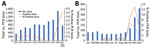
Figure 1. Positivity rate of Chlamydia pneumoniae PCRs in a tertiary care hospital, Lausanne, Switzerland. A) Yearly number of C. pneumoniaePCR tests conducted during 2014-2023. The final bar...
Figure 2
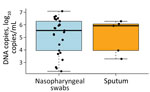
Figure 2. Boxplot of the quantifications of the Chlamydia pneumoniae–positive PCRs, by sample type, in a tertiary care hospital, Lausanne, Switzerland. In total, 24 nasopharyngeal swab and 5 sputum samples...
During routine epidemiologic surveillance at Lausanne University Hospital in Lausanne, Switzerland, positive C. pneumoniae bacteria PCR rates surged to 3.61% during October–December 2023, peaking at 6.66% in October, contrasting with the usual 0%–0.75% range reported over the past decade (Figure 1, panel A, B). The PCR method we used for testing has been previously described in Opota et al. (10). In this most recent outbreak, we documented C. pneumoniae bacteria in 28 patients in 2023; of those, 20 were children (mean age 8 years) and 8 were adults (mean age 43 years). Patients with C. pneumoniae bacteria sometimes reported wheezing as a major clinical complaint. We tested bacterial loads in patients positive for C. pneumoniae bacteria and found that the mean bacterial load was 1,534,821 DNA copies/mL (range 200–11,998,897 DNA copies/mL). We collected nasopharyngeal swabs most frequently (n = 24), whereas we collected sputum samples (n = 5) and nasal swab samples (nostril only, n = 1) less frequently. Of note, bacterial loads were not higher in the analyzed sputa than in the nasopharyngeal swabs (p = 1 by Wilcoxon rank-sum test) (Figure 2).
The results of this analysis should be interpreted with caution in the absence of a larger number of paired samples. This analysis includes only 2 paired samples exhibiting <1 logarithm (decimal) of difference in DNA copies per milliliter.
To explain this sudden surge of C. pneumoniae bacterial infection, we suspect 2 primary factors. First, decreased immunity may have developed because of fewer circulating strains in the population over the past 3 years, related to SARS-CoV-2 transmission prevention measures. Second, recently relaxed hygiene standards after the SARS-CoV-2 pandemic may have increased the risk for infection.
Clinical suspicion of C. pneumoniae infection is particularly warranted when patients’ clinical manifestations include a persistent dry cough or wheezing. Molecular testing, if available, should be the first-line diagnostic tool with nasopharyngeal swabs as an acceptable sample collection method. We do not recommend serologic testing in such cases because of the need to collect convalescent serum and the late appearance of antibodies. Antibodies generally develop 2–3 weeks after symptom onset for IgM and 4–8 weeks for IgG, which is rather late for diagnostic and therapeutic purposes. Furthermore, because C. pneumoniae bacterial infection can be treated by macrolides, doxycycline, or fluoroquinolones, current increases in both C. pneumoniae and M. pneumoniae bacteria lead us to recommend PCR testing for both bacteria in symptomatic patients, instead of testing only for respiratory viruses. Although co-infection with M. pneumoniae bacteria occurred in only 1 patient (M. pneumoniae PCR was tested on all samples) in our cohort, viral co-infections are not uncommon. Many multiplexed PCR respiratory panels are available and could help monitor the trend of C. pneumoniae bacterial infections on a larger scale.
In conclusion, we outline an upsurge of C. pneumoniae bacterial infections in the Lausanne region of Switzerland, especially in the pediatric population, raising concerns for other settings and regions. We found no clear epidemiologic link between patients, which suggests that we are detecting a minority of cases and that infections may occur at higher rates in the community than we have documented. This local finding highlights the importance of considering this intracellular bacterium as a causative agent, along with other fastidious organisms such as M. pneumoniae bacteria, which are also on the rise (9).
Dr. Tagini is a trainee in clinical microbiology and infectious diseases at the Lausanne University Hospital. His research interests are focused mainly on intracellular bacteria and bacterial genomics.
References
- Grayston JT, Campbell LA, Kuo CC, Mordhorst CH, Saikku P, Thom DH, et al. A new respiratory tract pathogen: Chlamydia pneumoniae strain TWAR. J Infect Dis. 1990;161:618–25. DOIPubMedGoogle Scholar
- Kumar S, Hammerschlag MR. Acute respiratory infection due to Chlamydia pneumoniae: current status of diagnostic methods. Clin Infect Dis. 2007;44:568–76. DOIPubMedGoogle Scholar
- Senn L, Jaton K, Fitting JW, Greub G. Does respiratory infection due to Chlamydia pneumoniae still exist? Clin Infect Dis. 2011;53:847–8. DOIPubMedGoogle Scholar
- Conklin L, Adjemian J, Loo J, Mandal S, Davis C, Parks S, et al. Investigation of a Chlamydia pneumoniae outbreak in a Federal correctional facility in Texas. Clin Infect Dis. 2013;57:639–47. DOIPubMedGoogle Scholar
- Han HY, Moon JU, Rhim JW, Kang HM, Lee SJ, Yang EA. Surge of Chlamydia pneumoniae pneumonia in children hospitalized with community-acquired pneumonia at a single center in korea in 2016. J Infect Chemother. 2023;29:453–7. DOIPubMedGoogle Scholar
- Hahn DL, Schure A, Patel K, Childs T, Drizik E, Webley W. Chlamydia pneumoniae-specific IgE is prevalent in asthma and is associated with disease severity. PLoS One. 2012;7:
e35945 . DOIPubMedGoogle Scholar - Pittet LF, Bertelli C, Scherz V, Rochat I, Mardegan C, Brouillet R, et al. Chlamydia pneumoniae and Mycoplasma pneumoniae in children with cystic fibrosis: impact on bacterial respiratory microbiota diversity. Pathog Dis. 2021;79:ftaa074.
- Meyer Sauteur PM, Beeton ML, Pereyre S, Bébéar C, Gardette M, Hénin N, et al.; ESGMAC the ESGMAC MAPS study group. Mycoplasma pneumoniae: gone forever? Lancet Microbe. 2023;4:
e763 . DOIPubMedGoogle Scholar - Meyer Sauteur PM, Beeton ML, Pereyre S, Bébéar C, Gardette M, Hénin N, et al.; European Society of Clinical Microbiology and Infectious Diseases (ESCMID) Study Group for Mycoplasma and Chlamydia Infections (ESGMAC), and the ESGMAC Mycoplasma pneumoniae Surveillance (MAPS) study group. Mycoplasma pneumoniae: delayed re-emergence after COVID-19 pandemic restrictions. Lancet Microbe. 2024;5:e100–1. DOIPubMedGoogle Scholar
- Opota O, Brouillet R, Greub G, Jaton K. Methods for real-time PCR-based diagnosis of Chlamydia pneumoniae, Chlamydia psittaci, and Chlamydia abortus infections in an opened molecular diagnostic platform. Methods Mol Biol. 2017;1616:171–81. DOIPubMedGoogle Scholar
Figures
Cite This ArticleOriginal Publication Date: February 27, 2024
Table of Contents – Volume 30, Number 4—April 2024
| EID Search Options |
|---|
|
|
|
|
|
|
Please use the form below to submit correspondence to the authors or contact them at the following address:
Gilbert Greub, Institute of Microbiology, Department of Laboratory Medicine and Pathology, Lausanne University Hospital and University of Lausanne, Bugnon 48, CH-1011 Lausanne, Switzerland
Top