Volume 6, Number 5—October 2000
Dispatch
Trichinella pseudospiralis Outbreak in France
Stéphane Ranque*, Bernard Faugère*†, Edoardo Pozio†, Giuseppe La Rosa†, Alessandra Tamburrini†, Jean-François Pellissier‡, and Philippe Brouqui*
Figure 1
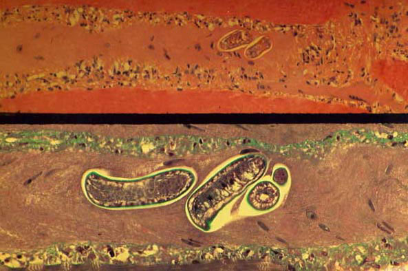
Figure 1. Sections of a muscle biopsy specimen from a patient infected with Trichinella pseudospiralis on day 32 after infection. The identified larva is nonencapsulated. Inflammatory cells are noted in the interstitium. Upper panel: hematoxylin and eosin stain at 50X magnification. Bottom panel: Masson trichrome stain at 100X magnification.
Page created: December 17, 2010
Page updated: December 17, 2010
Page reviewed: December 17, 2010
The conclusions, findings, and opinions expressed by authors contributing to this journal do not necessarily reflect the official position of the U.S. Department of Health and Human Services, the Public Health Service, the Centers for Disease Control and Prevention, or the authors' affiliated institutions. Use of trade names is for identification only and does not imply endorsement by any of the groups named above.