Volume 7, Number 5—October 2001
Dispatch
A Unique Mycobacterium Species Isolated from an Epizootic of Striped Bass (Morone saxatilis)
Abstract
We isolated a Mycobacterium sp. resembling Mycobacterium marinum and M. ulcerans from diseased striped bass (Morone saxatilis) during an epizootic of mycobacteriosis in the Chesapeake Bay. This isolate may represent an undescribed Mycobacterium species, based on phenotypic characteristics and comparative 16S rRNA gene sequence.
Natural aquatic environments are recognized sources of mycobacteria known to cause disease in both humans and fish. Although Mycobacterium marinum is considered the primary causative agent of fish mycobacteriosis, seven Mycobacterium species associated with tubercle granulomas in aquarium, cultured, and wild fish populations have been described: M. abscessus, M. chelonae, M. fortuitum, M. marinum, M. neoaurum, M. scrofulaceum, and M. simiae (1,2). All these species cause disease in humans (3,4). Primary clinical syndromes include skin and soft-tissue infections, cervical lymphadenitis, pulmonary disease, and disseminated infections, the last generally being limited to immunocompromised persons. Human mycobacteriosis following occupational or recreational exposure to the marine environment is frequently associated with trauma such as wounds from handling fish and has been attributed primarily to M. marinum (5). Consequently, the discovery of an undescribed Mycobacterium species associated with an epizootic of mycobacteriosis in striped bass (Morone saxatilis) warrants recognition and additional study.
Mycobacteriosis in fish is a subacute to chronic wasting disease known to affect some 167 freshwater and saltwater species (2). Internal signs of the disease vary according to fish species but typically include granulomas in the spleen, kidney, and liver. External manifestations include scale loss accompanied by hemorrhagic lesions penetrating the musculature in advanced cases. Recently, an ongoing epizootic of mycobacteriosis in striped bass (Morone saxatilis) from the Chesapeake Bay was described (Vogelbein W et al., unpub. data). Previous outbreaks of mycobacteriosis in wild striped bass have occurred in Pacific estuaries (6). During the Chesapeake Bay epizootic, we isolated a variety of mycobacteria associated with skin and visceral lesions that included a unique group of slowly growing nonpigmented isolates. We describe one of these isolates, which has specific characteristics similar to those of M. marinum and M. ulcerans.
Figure
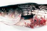
Figure. . Skin ulcers typical of mycobacteriosis in striped bass (Morone saxatilis) from the Chesapeake Bay.
Striped bass (n = 20) we examined included asymptomatic and symptomatic fish with skin ulcerations (Figure) verified histologically to exhibit granulomatous inflammation associated with acid-fast bacilli. All fish were caught in the Chesapeake Bay or one of its tributaries (the James, Potomac, or Rappahannock rivers). Skin and spleen samples from necropsied specimens were processed for routine paraffin histology, sectioned at 5 µm, and stained with hematoxylin and eosin. Selected sections were stained using Ziehl-Neelsen's method for acid-fast bacteria (7). Excised internal tissues (predominately spleen) were homogenized in phosphate buffer using a Ten Broeck tissue grinder and inoculated directly onto culture media or after treatment with one of the following disinfectants (Vogelbein et al., unpub. data): 0.3% Zephiran (Sanofi Winthrop Pharmaceuticals, New York, NY), 2% NaOH, or 2% HCl. Homogenates were inoculated onto Löwenstein-Jensen slants and plates of brain heart infusion agar containing 5% sheep red blood cells and Middlebrook 7H10 agar with albumin-dextrose-catalase enrichment. Initially inoculated media were incubated at 30°C for a minimum of 2 months. Because some isolates exhibited poor growth, a second incubation temperature (23°C) was used for primary media inoculated with tissue homogenates.
Purified isolates were characterized phenotypically by traditional methods (8) with incubation at 23°C. Mycolic acids were analyzed by a standardized method for mycobacteria by using reverse-phase high-performance liquid chromatography (HPLC) with UV detection (9,10).
Polymerase chain reaction (PCR) assay and sequence analysis of the 16S rRNA gene were used to characterize one of the slow-growing, nonpigmented mycobacteria, hereafter called isolate M175. This isolate is deposited in the American Type Culture Collection (ATCC), Rockville, MD, as ATCC 700981. The 16S rRNA gene was amplified in 120-µL volumes (11) by using cycle conditions described by van Berkum and Fuhrmann (12). Primers (forward, M16SA, 5'-CGC TGG CGG CGT GCT TA-3' and reverse, M16SB, 5'-ACG GCT ACC TTG TTA C-3') were specifically designed for the amplification of mycobacterial 16S rRNA genes. The PCR buffer (pH 8.5) contained 60 mM Tris-HCl, 15 mM (NH4)2SO4, and 1.5 mM MgCl2; control reactions without template were included. After purification of PCR products (QIAquick Spin columns, Qiagen Inc., Chatsworth, CA), amplicons were sequenced with a Perkin-Elmer 377 DNA Sequencer in combination with a Dye Deoxy Terminator Cycle Sequencing Kit (Perkin-Elmer, Foster City, CA) (11,12).
Granulomatous inflammation was confirmed histologically in spleens of 18 of the 20 fish. Severity of the infection based on the abundance and size of splenic granulomas varied from mild to severe. Skin ulcers were evident in 13 specimens. Granulomatous inflammation was generally associated with acid-fast bacilli in selected stained sections.
Colony development from homogenized tissue was slow, requiring 4 to 6 weeks' incubation at 23°C on the preferred medium, Middlebrook 7H10 agar. Isolate M175 showed little or no growth at 30°C and none at 37°C. Rough nonpigmented colonies were flat with an irregular margin and yielded aggregates of acid-fast nonbranching rods. Isolate M175 was negative for growth on MacConkey agar and Löwenstein-Jensen with 5% NaCl, arylsulfatase, beta-galactosidase, nitrate reductase, semiquantitative catalase, Tween 80 hydrolysis, and Tween opacity. Weak positive reactions for catalase activity after treatment at 68°C and pyrazinamidase after extending incubation to 14 days were observed. Isolate M175 was positive for tellurite reduction, niacin production, and urease. Colonies did not produce pigment after exposure to light for several hours or after prolonged exposure for several days. Based on the aforementioned characteristics, this isolate could not be assigned to an existing species.
The M175 mycolic acid pattern consisted of a single cluster of eight peaks that visually resembled reference patterns (10) for species of the M. tuberculosis complex. However, M175 mycolic acid peaks did not superimpose with peaks of M. tuberculosis after alignment with the internal size standard. Peak elution times for M175 were suggestive of more polar, shorter, carbon chain-length mycolic acids than those found in M. tuberculosis complex species. Comparisons of the M175 pattern with the Mycobacterium HPLC mycolic acid database at the Centers for Disease Control and Prevention confirmed a unique pattern suggestive of a new species of mycobacteria.
The sequence of the PCR product of the 16S rRNA gene from Mycobacterium isolate M175 was 1,494 nt long. This sequence was deposited in GenBank and was given accession number AY005147. Blast searches of GenBank yielded high sequence similarities of 99.2% to M. marinum (13) and M. ulcerans (14) and of 98.7% to M. bovis (15) and M. tuberculosis (16). High sequence similarities between 16S rRNA genes of M175 and other Mycobacterium spp. and phenotypic data support the conclusion that M175 belongs within the genus Mycobacterium. However, despite the similarities, the 16S gene sequence of M175 differed from M. ulcerans by 11 nt (3 insertions and 8 substitutions [one base of the M. ulcerans sequence in GenBank is N]) and from M. marinum by 10 nt (4 insertions, 6 substitutions [one base of the M. marinum sequence in GenBank is N]). Based on sequence differences and contrasting phenotypic characteristics (Table), we conclude that isolate M175 appears to belong to a new, previously undescribed species of Mycobacterium (19). Comparative genetic studies of M. ulcerans and M. marinum based on 16S rRNA sequence analysis have shown very close relationships between these species despite contrasting phenotypic profiles (20-24). The presence of two DNA insertion sequences, IS2404 and IS2606, in M. ulcerans but not in M. marinum has been used to distinguish the former (22-25).
The public health significance of this unique Mycobacterium species is not known. Frequently, mycobacterial disease in fish and cutaneous infections in humans are diagnosed on the basis of clinical presentation and generally attributed to M. marinum. Isolation of the causative agent either is not attempted or is unsuccessful, possibly because of loss of viability during specimen decontamination, inappropriate culture conditions, lack of technical experience with mycobacteria, or the prevailing assumption that detection of acid-fast rods is synonymous with a diagnosis of M. marinum. Consequently, the extent of environmentally acquired human infections caused by Mycobacterium species is not known. Studies to investigate the clinical importance of isolates obtained from persons exposed to marine or estuarine sources would provide data on which to evaluate the public health import of these isolates.
As in the present study, environmental mycobacteria may have lower temperature optima and not grow well on traditional media such as Löwenstein-Jensen. However, a preference for low temperature does not necessarily negate their ability to cause disease in humans, as demonstrated by disseminated infections caused by M. marinum and M. haemophilum or ulcerative skin disease caused by M. ulcerans. An epizootic of mycobacteriosis in striped bass, possibly the most important recreational fish in the Chesapeake Bay, could serve as a reservoir for transmission of mycobacterial infections to humans.
Laboratory challenge studies using striped bass are in progress to evaluate the pathogenicity of isolate M175. Additional research is needed to understand the persistence, distribution, and ecology of these mycobacterial isolates in natural waters, particularly with regard to their transmission to fish. Furthermore, this study also underlines a need to isolate and identify mycobacteria responsible for nontuberculosis infections in humans. This information is essential to determine the extent of human mycobacteriosis associated with occupational and increasingly popular recreational exposure to the natural aquatic environment.
Ms. Rhodes is a microbiologist in the Department of Environmental Sciences, Virginia Institute of Marine Science. Her research interests focus on public health microbiology related to the estuarine environment.
Acknowledgments
The authors thank Dana Booth, Dave Zwerner, and Patrick Elia for their excellent technical assistance.
Funding was obtained in part from the Virginia Marine Resource Commission, Commonwealth of Virginia, and the Virginia Institute of Marine Science, College of William and Mary (contribution no. 2368 of the Virginia Institute of Marine Science).
References
- Lansdell WB, Dixon B, Smith N, Benjamin L. Isolation of several Mycobacterium species from fish. J Aquat Anim Health. 1993;5:73–6. DOIGoogle Scholar
- Chinabut S. Mycobacteriosis and nocardiosis. In: Woo PTK, Bruno DW, editors. Fish diseases and disorders. Vol 3. Viral, bacterial and fungal infections. Wallington, UK: CAB International; 1999. p. 319-40.
- Wayne LG, Sramek HA. Agents of newly recognized or infrequently encountered mycobacterial diseases. Clin Microbiol Rev. 1992;5:1–25.PubMedGoogle Scholar
- Falkinham JO III. Epidemiology of infection by nontuberculous mycobacteria. Clin Microbiol Rev. 1996;9:177–215.PubMedGoogle Scholar
- Wolinsky E. Mycobacterial diseases other than tuberculosis. Clin Infect Dis. 1992;15:1–12. DOIPubMedGoogle Scholar
- Sakanari JA, Reilly CA, Moser M. Tubercular lesions in Pacific coast populations of striped bass. Trans Am Fish Soc. 1983;112:565–6. DOIGoogle Scholar
- Luna LG, ed. Manual of histologic staining methods of the Armed Forces Institute of Pathology. New York: McGraw-Hill; 1968.
- Lutz B. Section 3. Mycobacteriology. 3.12. Identification tests for mycobacteria. In: Isenburg HD, editor. Clinical microbiology procedures handbook. Vol 1. Washington: American Society for Microbiology; 1992. p. 3.12.1-29.
- Butler WR, Kilburn JO. Identification of major slowly growing pathogenic mycobacteria and Mycobacterium gordonae by high performance liquid chromatography of their mycolic acids. J Clin Microbiol. 1988;26:50–3.PubMedGoogle Scholar
- Butler WR, Floyd MM, Silcox V, Cage G, Desmond E, Duffey PS, Standardized method for HPLC identification of mycobacteria. HPLC users group in cooperation with Centers for Disease Control and Prevention. Atlanta: U.S. Public Health Service, CDC; 1996.
- van Berkum P, Beyene D, Eardly BD. Phylogenetic relationships among Rhizobium species nodulating the common bean Phaseolus vulgaris L. Int J Syst Bacteriol. 1996;46:240–4. DOIPubMedGoogle Scholar
- van Berkum P, Fuhrmann JJ. Evolutionary relationships among the soybean bradyrhizobia reconstructed from 16S rRNA gene and internally transcribed spacer region sequence divergence. Int J Syst Evol Microbiol. 2000;50:2165–72. DOIPubMedGoogle Scholar
- Rogall T, Wolters J, Flohr T, Bottger EC. Towards a phylogeny and definition of species at the molecular level within the genus Mycobacterium. Int J Syst Bacteriol. 1990;40:323–30. DOIPubMedGoogle Scholar
- Hofer M, Hirschel B, Kirschner P, Beghetti M, Kaelin A, Siegrist CA, Brief report: disseminated osteomyelitis from Mycobacterium ulcerans after a snakebite. N Engl J Med. 1993;328:1007–9. DOIPubMedGoogle Scholar
- Suzuki Y, Nagata A, Ono Y, Yamada T. Complete nucleotide sequence of the 16S rRNA gene of Mycobacterium bovis BCG. J Bacteriol. 1988;170:2886–9.PubMedGoogle Scholar
- Aranaz A, Liebana E, Gomez-Mampaso E, Galna JC, Cousins D, Ortega A, Mycobacterium tuberculosis subsp. caprae subsp. nov.: a taxonomic study of a new member of the Mycobacterium tuberculosis complex isolated from goats in Spain. Int J Syst Bacteriol. 1999;49:1263–73. DOIPubMedGoogle Scholar
- Witebsky FG, Kruczak-Filipov P. Identification of mycobacteria by conventional methods. Clin Lab Med. 1996;16:569–601.PubMedGoogle Scholar
- Goodfellow M, Magee JG. Taxonomy of mycobacteria. In: Gangadharam PRJ, Jenkins PA, editors. Mycobacteria I: Basic aspects. New York: Chapman and Hall; 1998. p. 1-71.
- Lévy-Frébault VV, Portaels F. Proposed minimal standards for the genus Mycobacterium and for description of new slowly growing Mycobacterium species. Int J Syst Bacteriol. 1992;42:315–23. DOIPubMedGoogle Scholar
- Portaels F, Fonteyne P-A, De Beenhouwer H, De Rijk P, Guédénon A, Hayman J, Variability in 3' end of 16S rRNA sequence of Mycobacterium ulcerans is related to geographic origin of isolates. J Clin Microbiol. 1996;34:962–5.PubMedGoogle Scholar
- Tonjum T, Welty DB, Jantzen E, Small PL. Differentiation of Mycobacterium ulcerans, M. marinum, and M. haemophilum: mapping of their relationships to M. tuberculosis by fatty acid profile analysis, DNA-DNA hybridization, and 16S rRNA gene sequence analysis. J Clin Microbiol. 1998;36:918–25.PubMedGoogle Scholar
- Stinear T, Ross BC, Davies JK, Marino L, Robins-Browne RM, Oppedisano F, Identification and characterization of IS2404 and IS2606: two distinct repeated sequences for detection of Mycobacterium ulcerans by PCR. J Clin Microbiol. 1999;37:1018–23.PubMedGoogle Scholar
- Stinear T, Davies JK, Jenkin GA, Portaels F, Ross BC, Oppedisano F, A simple PCR method for rapid genotype analysis of Mycobacterium ulcerans. J Clin Microbiol. 2000;38:11482–7.PubMedGoogle Scholar
- Stinear TP, Jenkin GA, Johnson PDR, Davies JK. Comparative genetic analysis of Mycobacterium ulcerans and Mycobacterium marinum reveals evidence of recent divergence. J Bacteriol. 2000;182:6322–30. DOIPubMedGoogle Scholar
- Ross BC, Marino L, Oppedisano F, Edwards R, Robins-Browne RM, Johnson PDR. Development of a PCR assay for rapid diagnosis of Mycobacteirum ulcerans infection. J Clin Microbiol. 1997;35:1696–700.PubMedGoogle Scholar
Figure
Table
Cite This ArticleTable of Contents – Volume 7, Number 5—October 2001
| EID Search Options |
|---|
|
|
|
|
|
|
Please use the form below to submit correspondence to the authors or contact them at the following address:
Martha W. Rhodes, Department of Environmental Sciences, Virginia Institute of Marine Science, College of William and Mary, PO Box 1346, Gloucester Point, VA 23062, USA; fax: 804-684-7186
Top