Volume 8, Number 4—April 2002
Synopsis
Baylisascaris procyonis: An Emerging Helminthic Zoonosis
Frank J. Sorvillo* , Lawrence R. Ash*, O.G.W. Berlin*†, JoAnne Yatabe†, Chris Degiorgio‡, and Stephen A. Morse§
Figure 1
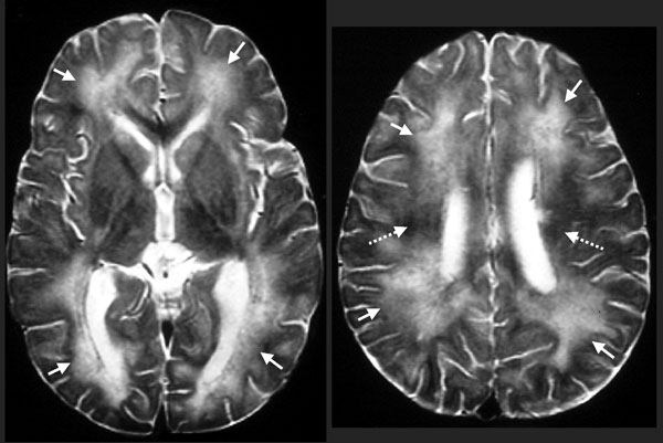
Figure 1. Biopsy-proven Baylisascaris procyonis encephalitis in a 13-month-old boy. Axial T2-weighted magnetic resonance images obtained 12 days after symptom onset show abnormal high signal throughout most of the central white matter (arrows) compared with the dark signal expected at this age (broken arrows).
Page created: July 15, 2010
Page updated: July 15, 2010
Page reviewed: July 15, 2010
The conclusions, findings, and opinions expressed by authors contributing to this journal do not necessarily reflect the official position of the U.S. Department of Health and Human Services, the Public Health Service, the Centers for Disease Control and Prevention, or the authors' affiliated institutions. Use of trade names is for identification only and does not imply endorsement by any of the groups named above.