Skip directly to site content Skip directly to page options Skip directly to A-Z link Skip directly to A-Z link Skip directly to A-Z link
Volume 19, Number 6—June 2013
Dispatch

BSE-associated Prion-Amyloid Cardiomyopathy in Primates

Susanne Krasemann, Giulia Mearini, Elisabeth Krämer, Katja Wagenführ, Walter Schulz-Schaeffer, Melanie Neumann, Walter Bodemer, Franz-Josef Kaup, Michael Beekes, Lucie Carrier, Adriano Aguzzi1, and Markus Glatzel1Comments to Author 
Author affiliations: University Medical Center Hamburg-Eppendorf, Hamburg, Germany (S. Krasemann, G. Mearini, E. Krämer, M. Neumann, L. Carrier, M. Glatzel); Robert Koch Institute, Berlin, Germany (K. Wagenführ, M. Beekes); University Hospital Göttingen, Göttingen, Germany (W. Schulz-Schaeffer); German Primate Center, Göttingen (W. Bodemer, F.-J. Kaup); University of Zurich, Zurich, Switzerland (A. Aguzzi)

Main Article

Figure 2

Abundant PrPSc in heart of 1 bovine spongiform encephalopathy (BSE)–infected rhesus macaque. A) In sodium phosphotungstic acid precipitation of PrPSc, followed by Western blotting, highly abundant PrPSc was demonstrated in the heart of 1 BSE-infected primate. In this monkey, only the heart contained PrPSc. Controls include cardiac muscle spiked with minimal amounts brain of a healthy (–) and prion-diseased (+) primate. All analyses were prepared from 50 mg of tissue except the heart of 1 monkey

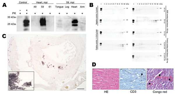
Figure 2. . Abundant PrPSc in heart of 1 bovine spongiform encephalopathy (BSE)–infected rhesus macaque. A) In sodium phosphotungstic acid precipitation of PrPSc, followed by Western blotting, highly abundant PrPSc was demonstrated in the heart of 1 BSE-infected primate. In this monkey, only the heart contained PrPSc. Controls include cardiac muscle spiked with minimal amounts brain of a healthy (–) and prion-diseased (+) primate. All analyses were prepared from 50 mg of tissue except the heart of 1 monkey 59 months postinoculation (mpi) (20 mg). PK, proteinase K. B) In protein-misfolding cyclic amplification, PrPSc was amplified only from the heart of 1 monkey 59 months postinoculation (mpi). As a positive control, brain tissue from a BSE-diseased monkey was used, and tissue from an uninfected control monkey served as a negative control. PK–digested hamster PrPSc (263 K) served as loading and digestion control for PrPSc. C) Paraffin-embedded tissue blotting of the entire heart of the 59 mpi monkey showed abundant deposition of PrPSc, mainly in the septum of the heart. Inset confirms the deposition pattern of PrPSc as amyloid. Scale bar = 0.25 mm. D) Histologic and immunohistochemical examination of heart tissue of the 59-mpi monkey by using hematoxylin and eosin (HE) staining and immunohistochemical staining against T-cell marker CD3 showed regularly configured cardiomyocytes and only single T-cells associated with blood vessels (arrow). Congo red staining showed Congo red–positive material in cardiomyocytes in a patch-like deposition pattern (arrows). Scale bar = 10 µm.

Main Article

1These authors contributed equally to this article.

Page created: May 20, 2013
Page updated: May 20, 2013
Page reviewed: May 20, 2013
The conclusions, findings, and opinions expressed by authors contributing to this journal do not necessarily reflect the official position of the U.S. Department of Health and Human Services, the Public Health Service, the Centers for Disease Control and Prevention, or the authors' affiliated institutions. Use of trade names is for identification only and does not imply endorsement by any of the groups named above.
file_external