Volume 22, Number 7—July 2016
Research
African Swine Fever Epidemic, Poland, 2014–2015
Krzysztof Śmietanka , Grzegorz Woźniakowski, Edyta Kozak, Krzysztof Niemczuk, Magdalena Frączyk, Łukasz Bocian, Andrzej Kowalczyk, and Zygmunt Pejsak
Figure 5
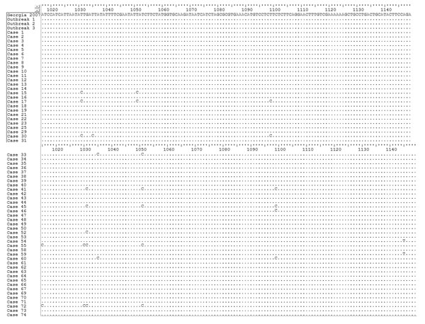
Figure 5. Nucleotide alignment of the MGF505–2R gene variable sequence fragment (residues from 1,015 to 1,149 nt) showing point mutations and differences between isolate Georgia 2007/1 isolate and African swine fever virus field isolates from Poland. The graph was generated by using Bioedit version 7.2.5 software (Ibis Biosciences, Carlsbad, CA, USA). The dots indicate identical nucleotide residues. The variable residues are visible as a nucleotide symbol.
Page created: June 14, 2016
Page updated: June 14, 2016
Page reviewed: June 14, 2016
The conclusions, findings, and opinions expressed by authors contributing to this journal do not necessarily reflect the official position of the U.S. Department of Health and Human Services, the Public Health Service, the Centers for Disease Control and Prevention, or the authors' affiliated institutions. Use of trade names is for identification only and does not imply endorsement by any of the groups named above.