Synopses
Turtle-Associated Salmonellosis, United States, 2006–2014 [PDF - 1.14 MB - 7 pages]
During 2006–2014, a total of 15 multistate outbreaks of turtle-associated salmonellosis in humans were reported in the United States. Exposure to small pet turtles has long been recognized as a source of human salmonellosis. The risk to public health has persisted and may be increasing. Turtles are a popular reptilian pet among children, and numerous risky behaviors for the zoonotic transmission of Salmonella bacteria to children have been reported in recent outbreaks. Despite a long-standing federal ban against the sale and distribution of turtles <4 in (<10.16 cm) long, these small reptiles can be readily acquired through multiple venues and continue to be the main source of turtle-associated salmonellosis in children. Enhanced efforts are needed to minimize the disease risk associated with small turtle exposure. Prevention will require novel partnerships and a comprehensive One Health approach involving human, animal, and environmental health.
| EID | Bosch S, Tauxe R, Behravesh C. Turtle-Associated Salmonellosis, United States, 2006–2014. Emerg Infect Dis. 2016;22(7):1149-1155. https://doi.org/10.3201/eid2207.150685 |
|---|---|
| AMA | Bosch S, Tauxe R, Behravesh C. Turtle-Associated Salmonellosis, United States, 2006–2014. Emerging Infectious Diseases. 2016;22(7):1149-1155. doi:10.3201/eid2207.150685. |
| APA | Bosch, S., Tauxe, R., & Behravesh, C. (2016). Turtle-Associated Salmonellosis, United States, 2006–2014. Emerging Infectious Diseases, 22(7), 1149-1155. https://doi.org/10.3201/eid2207.150685. |
Many of the survivors of the 2014–2015 epidemic of Ebola virus disease (EVD) in West Africa were women of childbearing age. Limited clinical and laboratory data exist that describe these women’s pregnancies and outcomes. We report the case of an EVD survivor who became pregnant and delivered her child in the United States, and we discuss implications of this case for infection control practices in obstetric services. Hospitals in the United States must be prepared to care for EVD survivors.
| EID | Kamali A, Jamieson DJ, Kpaduwa J, Schrier S, Kim M, Green NM, et al. Pregnancy, Labor, and Delivery after Ebola Virus Disease and Implications for Infection Control in Obstetric Services, United States. Emerg Infect Dis. 2016;22(7):1156-1161. https://doi.org/10.3201/eid2207.160269 |
|---|---|
| AMA | Kamali A, Jamieson DJ, Kpaduwa J, et al. Pregnancy, Labor, and Delivery after Ebola Virus Disease and Implications for Infection Control in Obstetric Services, United States. Emerging Infectious Diseases. 2016;22(7):1156-1161. doi:10.3201/eid2207.160269. |
| APA | Kamali, A., Jamieson, D. J., Kpaduwa, J., Schrier, S., Kim, M., Green, N. M....Mascola, L. (2016). Pregnancy, Labor, and Delivery after Ebola Virus Disease and Implications for Infection Control in Obstetric Services, United States. Emerging Infectious Diseases, 22(7), 1156-1161. https://doi.org/10.3201/eid2207.160269. |
Response to Emergence of Middle East Respiratory Syndrome Coronavirus, Abu Dhabi, United Arab Emirates, 2013–2014 [PDF - 495 KB - 7 pages]
In January 2013, several months after Middle East respiratory syndrome coronavirus (MERS-CoV) was first identified in Saudi Arabia, Abu Dhabi, United Arab Emirates, began surveillance for MERS-CoV. We analyzed medical chart and laboratory data collected by the Health Authority–Abu Dhabi during January 2013–May 2014. Using real-time reverse transcription PCR, we tested respiratory tract samples for MERS-CoV and identified 65 case-patients. Of these patients, 23 (35%) were asymptomatic at the time of testing, and 4 (6%) showed positive test results for >3 weeks (1 had severe symptoms and 3 had mild symptoms). We also identified 6 clusters of MERS-CoV cases. This report highlights the potential for virus shedding by mildly ill and asymptomatic case-patients. These findings will be useful for MERS-CoV management and infection prevention strategies.
| EID | Al Hosani F, Pringle K, Al Mulla M, Kim L, Pham HT, Alami NN, et al. Response to Emergence of Middle East Respiratory Syndrome Coronavirus, Abu Dhabi, United Arab Emirates, 2013–2014. Emerg Infect Dis. 2016;22(7):1162-1168. https://doi.org/10.3201/eid2207.160040 |
|---|---|
| AMA | Al Hosani F, Pringle K, Al Mulla M, et al. Response to Emergence of Middle East Respiratory Syndrome Coronavirus, Abu Dhabi, United Arab Emirates, 2013–2014. Emerging Infectious Diseases. 2016;22(7):1162-1168. doi:10.3201/eid2207.160040. |
| APA | Al Hosani, F., Pringle, K., Al Mulla, M., Kim, L., Pham, H. T., Alami, N. N....Gerber, S. I. (2016). Response to Emergence of Middle East Respiratory Syndrome Coronavirus, Abu Dhabi, United Arab Emirates, 2013–2014. Emerging Infectious Diseases, 22(7), 1162-1168. https://doi.org/10.3201/eid2207.160040. |
In the United States, Lyme disease is caused by Borrelia burgdorferi and transmitted to humans by blacklegged ticks. Patients with an erythema migrans lesion and epidemiologic risk can receive a diagnosis without laboratory testing. For all other patients, laboratory testing is necessary to confirm the diagnosis, but proper interpretation depends on symptoms and timing of illness. The recommended laboratory test in the United States is 2-tiered serologic analysis consisting of an enzyme-linked immunoassay or immunofluorescence assay, followed by reflexive immunoblotting. Sensitivity of 2-tiered testing is low (30%–40%) during early infection while the antibody response is developing (window period). For disseminated Lyme disease, sensitivity is 70%–100%. Specificity is high (>95%) during all stages of disease. Use of other diagnostic tests for Lyme disease is limited. We review the rationale behind current US testing guidelines, appropriate use and interpretation of tests, and recent developments in Lyme disease diagnostics.
| EID | Moore A, Nelson CA, Molins C, Mead PS, Schriefer M. Current Guidelines, Common Clinical Pitfalls, and Future Directions for Laboratory Diagnosis of Lyme Disease, United States. Emerg Infect Dis. 2016;22(7):1169-1177. https://doi.org/10.3201/eid2207.151694 |
|---|---|
| AMA | Moore A, Nelson CA, Molins C, et al. Current Guidelines, Common Clinical Pitfalls, and Future Directions for Laboratory Diagnosis of Lyme Disease, United States. Emerging Infectious Diseases. 2016;22(7):1169-1177. doi:10.3201/eid2207.151694. |
| APA | Moore, A., Nelson, C. A., Molins, C., Mead, P. S., & Schriefer, M. (2016). Current Guidelines, Common Clinical Pitfalls, and Future Directions for Laboratory Diagnosis of Lyme Disease, United States. Emerging Infectious Diseases, 22(7), 1169-1177. https://doi.org/10.3201/eid2207.151694. |
Two Linked Enteroinvasive Escherichia coli Outbreaks, Nottingham, UK, June 2014 [PDF - 823 KB - 7 pages]
Enteroinvasive Escherichia coli (EIEC) outbreaks are uncommon in Europe. In June 2014, two EIEC outbreaks occurred in Nottingham, UK, within 2 days; outbreak A was linked to a takeaway restaurant and outbreak B to a wedding party. We conducted 2 analytical studies: a case–control study for outbreak A and a cohort study for outbreak B. We tested microbiological and environmental samples, including by using whole-genome sequencing. For both outbreaks combined, we identified 157 probable case-patients; 27 were laboratory-confirmed as EIEC O96:H19–positive. Combined epidemiologic, microbiological, and environmental findings implicated lettuce as the vehicle of infection in outbreak A, but the source of the organism remained unknown. Whole-genome sequencing identified the same organism in cases from both outbreaks, but no epidemiologic link was confirmed. These outbreaks highlight that EIEC has the capacity to cause large and severe gastrointestinal disease outbreaks and should be considered as a potential pathogen in foodborne outbreaks in Europe.
| EID | Newitt S, MacGregor V, Robbins V, Bayliss L, Chattaway M, Dallman T, et al. Two Linked Enteroinvasive Escherichia coli Outbreaks, Nottingham, UK, June 2014. Emerg Infect Dis. 2016;22(7):1178-1184. https://doi.org/10.3201/eid2207.152080 |
|---|---|
| AMA | Newitt S, MacGregor V, Robbins V, et al. Two Linked Enteroinvasive Escherichia coli Outbreaks, Nottingham, UK, June 2014. Emerging Infectious Diseases. 2016;22(7):1178-1184. doi:10.3201/eid2207.152080. |
| APA | Newitt, S., MacGregor, V., Robbins, V., Bayliss, L., Chattaway, M., Dallman, T....Hawker, J. (2016). Two Linked Enteroinvasive Escherichia coli Outbreaks, Nottingham, UK, June 2014. Emerging Infectious Diseases, 22(7), 1178-1184. https://doi.org/10.3201/eid2207.152080. |
A Literature Review of Zika Virus [PDF - 800 KB - 8 pages]
Zika virus is a mosquitoborne flavivirus that is the focus of an ongoing pandemic and public health emergency. Previously limited to sporadic cases in Africa and Asia, the emergence of Zika virus in Brazil in 2015 heralded rapid spread throughout the Americas. Although most Zika virus infections are characterized by subclinical or mild influenza-like illness, severe manifestations have been described, including Guillain-Barre syndrome in adults and microcephaly in babies born to infected mothers. Neither an effective treatment nor a vaccine is available for Zika virus; therefore, the public health response primarily focuses on preventing infection, particularly in pregnant women. Despite growing knowledge about this virus, questions remain regarding the virus’s vectors and reservoirs, pathogenesis, genetic diversity, and potential synergistic effects of co-infection with other circulating viruses. These questions highlight the need for research to optimize surveillance, patient management, and public health intervention in the current Zika virus epidemic.
| EID | Plourde AR, Bloch EM. A Literature Review of Zika Virus. Emerg Infect Dis. 2016;22(7):1185-1192. https://doi.org/10.3201/eid2207.151990 |
|---|---|
| AMA | Plourde AR, Bloch EM. A Literature Review of Zika Virus. Emerging Infectious Diseases. 2016;22(7):1185-1192. doi:10.3201/eid2207.151990. |
| APA | Plourde, A. R., & Bloch, E. M. (2016). A Literature Review of Zika Virus. Emerging Infectious Diseases, 22(7), 1185-1192. https://doi.org/10.3201/eid2207.151990. |
Research
Comparing Characteristics of Sporadic and Outbreak-Associated Foodborne Illnesses, United States, 2004–2011 [PDF - 2.36 MB - 8 pages]
Outbreak data have been used to estimate the proportion of illnesses attributable to different foods. Applying outbreak-based attribution estimates to nonoutbreak foodborne illnesses requires an assumption of similar exposure pathways for outbreak and sporadic illnesses. This assumption cannot be tested, but other comparisons can assess its veracity. Our study compares demographic, clinical, temporal, and geographic characteristics of outbreak and sporadic illnesses from Campylobacter, Escherichia coli O157, Listeria, and Salmonella bacteria ascertained by the Foodborne Diseases Active Surveillance Network (FoodNet). Differences among FoodNet sites in outbreak and sporadic illnesses might reflect differences in surveillance practices. For Campylobacter, Listeria, and Escherichia coli O157, outbreak and sporadic illnesses are similar for severity, sex, and age. For Salmonella, outbreak and sporadic illnesses are similar for severity and sex. Nevertheless, the percentage of outbreak illnesses in the youngest age category was lower. Therefore, we do not reject the assumption that outbreak and sporadic illnesses are similar.
| EID | Ebel ED, Williams MS, Cole D, Travis CC, Klontz KC, Golden NJ, et al. Comparing Characteristics of Sporadic and Outbreak-Associated Foodborne Illnesses, United States, 2004–2011. Emerg Infect Dis. 2016;22(7):1193-1200. https://doi.org/10.3201/eid2207.150833 |
|---|---|
| AMA | Ebel ED, Williams MS, Cole D, et al. Comparing Characteristics of Sporadic and Outbreak-Associated Foodborne Illnesses, United States, 2004–2011. Emerging Infectious Diseases. 2016;22(7):1193-1200. doi:10.3201/eid2207.150833. |
| APA | Ebel, E. D., Williams, M. S., Cole, D., Travis, C. C., Klontz, K. C., Golden, N. J....Hoekstra, R. M. (2016). Comparing Characteristics of Sporadic and Outbreak-Associated Foodborne Illnesses, United States, 2004–2011. Emerging Infectious Diseases, 22(7), 1193-1200. https://doi.org/10.3201/eid2207.150833. |
African Swine Fever Epidemic, Poland, 2014–2015 [PDF - 2.32 MB - 7 pages]
In Poland, African swine fever (ASF) emerged in February 2014; by August 2015, the virus had been detected in >130 wild boar and in pigs in 3 backyard holdings. We evaluated ASF spread in Poland during these 18 months. Phylogenetic analysis indicated repeated incursions of genetically distinct ASF viruses of genotype II; the number of cases positively correlated wild boar density; and disease spread was very slow. More cases were reported during summer than autumn. The 18-month prevalence of ASF in areas under various animal movement restrictions was 18.6% among wild boar found dead or killed by vehicles and only 0.2% in hunted wild boar. Repeated introductions of the virus into the country, the primary role of wild boar in virus maintenance, and the slow spread of the disease indicate a need for enhanced biosecurity at pig holdings and continuous and intensive surveillance for fast detection of ASF.
| EID | Śmietanka K, Woźniakowski G, Kozak E, Niemczuk K, Frączyk M, Bocian Ł, et al. African Swine Fever Epidemic, Poland, 2014–2015. Emerg Infect Dis. 2016;22(7):1201-1207. https://doi.org/10.3201/eid2207.151708 |
|---|---|
| AMA | Śmietanka K, Woźniakowski G, Kozak E, et al. African Swine Fever Epidemic, Poland, 2014–2015. Emerging Infectious Diseases. 2016;22(7):1201-1207. doi:10.3201/eid2207.151708. |
| APA | Śmietanka, K., Woźniakowski, G., Kozak, E., Niemczuk, K., Frączyk, M., Bocian, Ł....Pejsak, Z. (2016). African Swine Fever Epidemic, Poland, 2014–2015. Emerging Infectious Diseases, 22(7), 1201-1207. https://doi.org/10.3201/eid2207.151708. |
Restaurant Cooking Trends and Increased Risk for Campylobacter Infection [PDF - 1.29 MB - 8 pages]
In the United Kingdom, outbreaks of Campylobacter infection are increasingly attributed to undercooked chicken livers, yet many recipes, including those of top chefs, advocate short cooking times and serving livers pink. During 2015, we studied preferences of chefs and the public in the United Kingdom and investigated the link between liver rareness and survival of Campylobacter. We used photographs to assess chefs’ ability to identify chicken livers meeting safe cooking guidelines. To investigate the microbiological safety of livers chefs preferred to serve, we modeled Campylobacter survival in infected chicken livers cooked to various temperatures. Most chefs correctly identified safely cooked livers but overestimated the public’s preference for rareness and thus preferred to serve them more rare. We estimated that 19%–52% of livers served commercially in the United Kingdom fail to reach 70°C and that predicted Campylobacter survival rates are 48%–98%. These findings indicate that cooking trends are linked to increasing Campylobacter infections.
| EID | Jones AK, Rigby D, Burton M, Millman C, Williams NJ, Jones TR, et al. Restaurant Cooking Trends and Increased Risk for Campylobacter Infection. Emerg Infect Dis. 2016;22(7):1208-1215. https://doi.org/10.3201/eid2207.151775 |
|---|---|
| AMA | Jones AK, Rigby D, Burton M, et al. Restaurant Cooking Trends and Increased Risk for Campylobacter Infection. Emerging Infectious Diseases. 2016;22(7):1208-1215. doi:10.3201/eid2207.151775. |
| APA | Jones, A. K., Rigby, D., Burton, M., Millman, C., Williams, N. J., Jones, T. R....Cross, P. (2016). Restaurant Cooking Trends and Increased Risk for Campylobacter Infection. Emerging Infectious Diseases, 22(7), 1208-1215. https://doi.org/10.3201/eid2207.151775. |
Heat Wave–Associated Vibriosis, Sweden and Finland, 2014 [PDF - 2.33 MB - 5 pages]
During summer 2014, a total of 89 Vibrio infections were reported in Sweden and Finland, substantially more yearly infections than previously have been reported in northern Europe. Infections were spread across most coastal counties of Sweden and Finland, but unusually, numerous infections were reported in subarctic regions; cases were reported as far north as 65°N, ≈100 miles (160 km) from the Arctic Circle. Most infections were caused by non-O1/O139 V. cholerae (70 cases, corresponding to 77% of the total, all strains were negative for the cholera toxin gene). An extreme heat wave in northern Scandinavia during summer 2014 led to unprecedented high sea surface temperatures, which appear to have been responsible for the emergence of Vibrio bacteria at these latitudes. The emergence of vibriosis in high-latitude regions requires improved diagnostic detection and clinical awareness of these emerging pathogens.
| EID | Baker-Austin C, Trinanes J, Salmenlinna S, Löfdahl M, Siitonen A, Taylor N, et al. Heat Wave–Associated Vibriosis, Sweden and Finland, 2014. Emerg Infect Dis. 2016;22(7):1216-1220. https://doi.org/10.3201/eid2207.151996 |
|---|---|
| AMA | Baker-Austin C, Trinanes J, Salmenlinna S, et al. Heat Wave–Associated Vibriosis, Sweden and Finland, 2014. Emerging Infectious Diseases. 2016;22(7):1216-1220. doi:10.3201/eid2207.151996. |
| APA | Baker-Austin, C., Trinanes, J., Salmenlinna, S., Löfdahl, M., Siitonen, A., Taylor, N....Martinez-Urtaza, J. (2016). Heat Wave–Associated Vibriosis, Sweden and Finland, 2014. Emerging Infectious Diseases, 22(7), 1216-1220. https://doi.org/10.3201/eid2207.151996. |
High Incidence of Chikungunya Virus and Frequency of Viremic Blood Donations during Epidemic, Puerto Rico, USA, 2014 [PDF - 718 KB - 8 pages]
Chikungunya virus (CHIKV) caused large epidemics throughout the Caribbean in 2014. We conducted nucleic acid amplification testing (NAAT) for CHIKV RNA (n = 29,695) and serologic testing for IgG against CHIKV (n = 1,232) in archived blood donor samples collected during and after an epidemic in Puerto Rico in 2014. NAAT yields peaked in October with 2.1% of donations positive for CHIKV RNA. A total of 14% of NAAT-reactive donations posed a high risk for virus transmission by transfusion because of high virus RNA copy numbers (104–109 RNA copies/mL) and a lack of specific IgM and IgG responses. Testing of minipools of 16 donations would not have detected 62.5% of RNA-positive donations detectable by individual donor testing, including individual donations without IgM and IgG. Serosurveys before and after the epidemic demonstrated that nearly 25% of blood donors in Puerto Rico acquired CHIKV infections and seroconverted during the epidemic.
| EID | Simmons G, Brès V, Lu K, Liss NM, Brambilla DJ, Ryff KR, et al. High Incidence of Chikungunya Virus and Frequency of Viremic Blood Donations during Epidemic, Puerto Rico, USA, 2014. Emerg Infect Dis. 2016;22(7):1221-1228. https://doi.org/10.3201/eid2207.160116 |
|---|---|
| AMA | Simmons G, Brès V, Lu K, et al. High Incidence of Chikungunya Virus and Frequency of Viremic Blood Donations during Epidemic, Puerto Rico, USA, 2014. Emerging Infectious Diseases. 2016;22(7):1221-1228. doi:10.3201/eid2207.160116. |
| APA | Simmons, G., Brès, V., Lu, K., Liss, N. M., Brambilla, D. J., Ryff, K. R....Murphy, E. L. (2016). High Incidence of Chikungunya Virus and Frequency of Viremic Blood Donations during Epidemic, Puerto Rico, USA, 2014. Emerging Infectious Diseases, 22(7), 1221-1228. https://doi.org/10.3201/eid2207.160116. |
Tropheryma whipplei as a Cause of Epidemic Fever, Senegal, 2010–2012 [PDF - 560 KB - 6 pages]
The bacterium Tropheryma whipplei, which causes Whipple disease in humans, is commonly detected in the feces of persons in Africa. It is also associated with acute infections. We investigated the role of T. whipplei in febrile patients from 2 rural villages in Senegal. During June 2010–March 2012, we collected whole-blood finger-prick samples from 786 febrile and 385 healthy villagers. T. whipplei was detected in blood specimens from 36 (4.6%) of the 786 febrile patients and in 1 (0.25%) of the 385 apparently healthy persons. Of the 37 T. whipplei cases, 26 (70.2%) were detected in August 2010. Familial cases and a potential new genotype were observed. The patients’ symptoms were mainly headache (68.9%) and cough (36.1%). Our findings suggest that T. whipplei is a cause of epidemic fever in Senegal.
| EID | Bassene H, Mediannikov O, Socolovschi C, Ratmanov P, Keita AK, Sokhna C, et al. Tropheryma whipplei as a Cause of Epidemic Fever, Senegal, 2010–2012. Emerg Infect Dis. 2016;22(7):1229-1334. https://doi.org/10.3201/eid2207.150441 |
|---|---|
| AMA | Bassene H, Mediannikov O, Socolovschi C, et al. Tropheryma whipplei as a Cause of Epidemic Fever, Senegal, 2010–2012. Emerging Infectious Diseases. 2016;22(7):1229-1334. doi:10.3201/eid2207.150441. |
| APA | Bassene, H., Mediannikov, O., Socolovschi, C., Ratmanov, P., Keita, A. K., Sokhna, C....Fenollar, F. (2016). Tropheryma whipplei as a Cause of Epidemic Fever, Senegal, 2010–2012. Emerging Infectious Diseases, 22(7), 1229-1334. https://doi.org/10.3201/eid2207.150441. |
Dispatches
Outbreak of Vibrio parahaemolyticus Sequence Type 120, Peru, 2009 [PDF - 535 KB - 3 pages]
In 2009, an outbreak of Vibrio parahaemolyticus occurred in Piura, Cajamarca, Lambayeque, and Lima, Peru. Whole-genome sequencing of clinical and environmental samples from the outbreak revealed a new V. parahaemolyticus clone. All the isolates identified belonged to a single clonal complex described exclusively in Asia before its emergence in Peru.
| EID | Gonzalez-Escalona N, Gavilan RG, Toro M, Zamudio ML, Martinez-Urtaza J. Outbreak of Vibrio parahaemolyticus Sequence Type 120, Peru, 2009. Emerg Infect Dis. 2016;22(7):1235-1237. https://doi.org/10.3201/eid2207.151896 |
|---|---|
| AMA | Gonzalez-Escalona N, Gavilan RG, Toro M, et al. Outbreak of Vibrio parahaemolyticus Sequence Type 120, Peru, 2009. Emerging Infectious Diseases. 2016;22(7):1235-1237. doi:10.3201/eid2207.151896. |
| APA | Gonzalez-Escalona, N., Gavilan, R. G., Toro, M., Zamudio, M. L., & Martinez-Urtaza, J. (2016). Outbreak of Vibrio parahaemolyticus Sequence Type 120, Peru, 2009. Emerging Infectious Diseases, 22(7), 1235-1237. https://doi.org/10.3201/eid2207.151896. |
Clinical Manifestations of Senecavirus A Infection in Neonatal Pigs, Brazil, 2015 [PDF - 1.44 MB - 4 pages]
We identified new clinical manifestations associated with Senecavirus A infection in neonatal piglets in Brazil in 2015. Immunohistochemical and molecular findings confirmed the association of Senecavirus A with these unusual clinical signs and more deaths. Other possible disease agents investigated were not associated with these illnesses.
| EID | Leme RA, Oliveira T, Alcântara BK, Headley SA, Alfieri AF, Yang M, et al. Clinical Manifestations of Senecavirus A Infection in Neonatal Pigs, Brazil, 2015. Emerg Infect Dis. 2016;22(7):1238-1241. https://doi.org/10.3201/eid2207.151583 |
|---|---|
| AMA | Leme RA, Oliveira T, Alcântara BK, et al. Clinical Manifestations of Senecavirus A Infection in Neonatal Pigs, Brazil, 2015. Emerging Infectious Diseases. 2016;22(7):1238-1241. doi:10.3201/eid2207.151583. |
| APA | Leme, R. A., Oliveira, T., Alcântara, B. K., Headley, S. A., Alfieri, A. F., Yang, M....Alfieri, A. A. (2016). Clinical Manifestations of Senecavirus A Infection in Neonatal Pigs, Brazil, 2015. Emerging Infectious Diseases, 22(7), 1238-1241. https://doi.org/10.3201/eid2207.151583. |
Infection with Possible Novel Parapoxvirus in Horse, Finland, 2013 [PDF - 1.65 MB - 4 pages]
A horse in Finland exhibited generalized granulomatous inflammation and severe proliferative dermatitis. After euthanization, we detected poxvirus DNA from a skin lesion sample. The virus sequence grouped with parapoxviruses, closely resembling a novel poxvirus detected in humans in the United States after horse contact. Our findings indicate horses may be a reservoir for zoonotic parapoxvirus.
| EID | Airas N, Hautaniemi M, Syrjä P, Knuuttila A, Putkuri N, Coulter L, et al. Infection with Possible Novel Parapoxvirus in Horse, Finland, 2013. Emerg Infect Dis. 2016;22(7):1242-1245. https://doi.org/10.3201/eid2207.151636 |
|---|---|
| AMA | Airas N, Hautaniemi M, Syrjä P, et al. Infection with Possible Novel Parapoxvirus in Horse, Finland, 2013. Emerging Infectious Diseases. 2016;22(7):1242-1245. doi:10.3201/eid2207.151636. |
| APA | Airas, N., Hautaniemi, M., Syrjä, P., Knuuttila, A., Putkuri, N., Coulter, L....Kinnunen, P. M. (2016). Infection with Possible Novel Parapoxvirus in Horse, Finland, 2013. Emerging Infectious Diseases, 22(7), 1242-1245. https://doi.org/10.3201/eid2207.151636. |
Vesicular Disease in 9-Week-Old Pigs Experimentally Infected with Senecavirus A [PDF - 1.15 MB - 3 pages]
Senecavirus A has been infrequently associated with vesicular disease in swine since 1988. However, clinical disease has not been reproduced after experimental infection with this virus. We report vesicular disease in 9-week-old pigs after Sencavirus A infection by the intranasal route under experimental conditions.
| EID | Montiel N, Buckley A, Guo B, Kulshreshtha V, VanGeelen A, Hoang H, et al. Vesicular Disease in 9-Week-Old Pigs Experimentally Infected with Senecavirus A. Emerg Infect Dis. 2016;22(7):1246-1248. https://doi.org/10.3201/eid2207.151863 |
|---|---|
| AMA | Montiel N, Buckley A, Guo B, et al. Vesicular Disease in 9-Week-Old Pigs Experimentally Infected with Senecavirus A. Emerging Infectious Diseases. 2016;22(7):1246-1248. doi:10.3201/eid2207.151863. |
| APA | Montiel, N., Buckley, A., Guo, B., Kulshreshtha, V., VanGeelen, A., Hoang, H....Lager, K. (2016). Vesicular Disease in 9-Week-Old Pigs Experimentally Infected with Senecavirus A. Emerging Infectious Diseases, 22(7), 1246-1248. https://doi.org/10.3201/eid2207.151863. |
Hepatitis E Virus Infection in Dromedaries, North and East Africa, United Arab Emirates, and Pakistan, 1983–2015 [PDF - 1.53 MB - 4 pages]
A new hepatitis E virus (HEV-7) was recently found in dromedaries and 1 human from the United Arab Emirates. We screened 2,438 dromedary samples from Pakistan, the United Arab Emirates, and 4 African countries. HEV-7 is long established, diversified and geographically widespread. Dromedaries may constitute a neglected source of zoonotic HEV infections.
| EID | Rasche A, Saqib M, Liljander AM, Bornstein S, Zohaib A, Renneker S, et al. Hepatitis E Virus Infection in Dromedaries, North and East Africa, United Arab Emirates, and Pakistan, 1983–2015. Emerg Infect Dis. 2016;22(7):1249-1252. https://doi.org/10.3201/eid2207.160168 |
|---|---|
| AMA | Rasche A, Saqib M, Liljander AM, et al. Hepatitis E Virus Infection in Dromedaries, North and East Africa, United Arab Emirates, and Pakistan, 1983–2015. Emerging Infectious Diseases. 2016;22(7):1249-1252. doi:10.3201/eid2207.160168. |
| APA | Rasche, A., Saqib, M., Liljander, A. M., Bornstein, S., Zohaib, A., Renneker, S....Corman, V. (2016). Hepatitis E Virus Infection in Dromedaries, North and East Africa, United Arab Emirates, and Pakistan, 1983–2015. Emerging Infectious Diseases, 22(7), 1249-1252. https://doi.org/10.3201/eid2207.160168. |
Increased Mortality Rates Associated with Staphylococcus aureus and Influenza Co-infection, Maryland and Iowa, USA [PDF - 985 KB - 4 pages]
We retrospectively analyzed data for 195 respiratory infection patients who had positive Staphyloccocus aureus cultures and who were hospitalized in 2 hospitals in Iowa and Maryland, USA, during 2003–2009. Odds for death for patients who also had influenza-positive test results were >4 times higher than for those who had negative influenza test results.
| EID | McDanel JS, Perencevich EN, Storm J, Diekema DJ, Herwaldt L, Johnson J, et al. Increased Mortality Rates Associated with Staphylococcus aureus and Influenza Co-infection, Maryland and Iowa, USA. Emerg Infect Dis. 2016;22(7):1253-1256. https://doi.org/10.3201/eid2207.151319 |
|---|---|
| AMA | McDanel JS, Perencevich EN, Storm J, et al. Increased Mortality Rates Associated with Staphylococcus aureus and Influenza Co-infection, Maryland and Iowa, USA. Emerging Infectious Diseases. 2016;22(7):1253-1256. doi:10.3201/eid2207.151319. |
| APA | McDanel, J. S., Perencevich, E. N., Storm, J., Diekema, D. J., Herwaldt, L., Johnson, J....Schweizer, M. L. (2016). Increased Mortality Rates Associated with Staphylococcus aureus and Influenza Co-infection, Maryland and Iowa, USA. Emerging Infectious Diseases, 22(7), 1253-1256. https://doi.org/10.3201/eid2207.151319. |
Extended-Spectrum Cephalosporin-Resistant Salmonella enterica serovar Heidelberg Strains, the Netherlands [PDF - 1.99 MB - 5 pages]
Extended-spectrum cephalosporin-resistant Salmonella enterica serovar Heidelberg strains (JF6X01.0022/XbaI.0251, JF6X01.0326/XbaI.1966, JF6X01.0258/XbaI.1968, and JF6X01.0045/XbaI.1970) have been identified in the United States with pulsed-field gel electrophoresis. Our examination of isolates showed introduction of these strains in the Netherlands and highlight the need for active surveillance and intervention strategies by public health organizations.
| EID | Liakopoulos A, Geurts Y, Dierikx CM, Brouwer M, Kant A, Wit B, et al. Extended-Spectrum Cephalosporin-Resistant Salmonella enterica serovar Heidelberg Strains, the Netherlands. Emerg Infect Dis. 2016;22(7):1257-1261. https://doi.org/10.3201/eid2207.151377 |
|---|---|
| AMA | Liakopoulos A, Geurts Y, Dierikx CM, et al. Extended-Spectrum Cephalosporin-Resistant Salmonella enterica serovar Heidelberg Strains, the Netherlands. Emerging Infectious Diseases. 2016;22(7):1257-1261. doi:10.3201/eid2207.151377. |
| APA | Liakopoulos, A., Geurts, Y., Dierikx, C. M., Brouwer, M., Kant, A., Wit, B....Mevius, D. J. (2016). Extended-Spectrum Cephalosporin-Resistant Salmonella enterica serovar Heidelberg Strains, the Netherlands. Emerging Infectious Diseases, 22(7), 1257-1261. https://doi.org/10.3201/eid2207.151377. |
Identification of Streptococcus suis Meningitis through Population-Based Surveillance, Togo, 2010–2014 [PDF - 403 KB - 3 pages]
During 2010–2014, we enrolled 511 patients with suspected bacterial meningitis into surveillance in 2 districts of northern Togo. We identified 15 persons with Streptococcus suis infection; 10 had occupational contact with pigs, and 12 suffered neurologic sequelae. S. suis testing should be considered in rural areas of the African meningitis belt.
| EID | Tall H, Njanpop-Lafourcade B, Mounkoro D, Tidjani L, Agbenoko K, Alassani I, et al. Identification of Streptococcus suis Meningitis through Population-Based Surveillance, Togo, 2010–2014. Emerg Infect Dis. 2016;22(7):1262-1264. https://doi.org/10.3201/eid2207.151511 |
|---|---|
| AMA | Tall H, Njanpop-Lafourcade B, Mounkoro D, et al. Identification of Streptococcus suis Meningitis through Population-Based Surveillance, Togo, 2010–2014. Emerging Infectious Diseases. 2016;22(7):1262-1264. doi:10.3201/eid2207.151511. |
| APA | Tall, H., Njanpop-Lafourcade, B., Mounkoro, D., Tidjani, L., Agbenoko, K., Alassani, I....Moïsi, J. C. (2016). Identification of Streptococcus suis Meningitis through Population-Based Surveillance, Togo, 2010–2014. Emerging Infectious Diseases, 22(7), 1262-1264. https://doi.org/10.3201/eid2207.151511. |
Postbooster Antibodies from Humans as Source of Diphtheria Antitoxin [PDF - 304 KB - 3 pages]
Diphtheria antitoxin for therapeutic use is in limited supply. A potential source might be affinity-purified antibodies originally derived from plasma of adults who received a booster dose of a vaccine containing diphtheria toxoid. These antibodies might be useful for treating even severe cases of diphtheria.
| EID | Bermejo-Martin JF, Avila-Alonso A, González-Rivera M, Tamayo E, Eiros J, Almansa R. Postbooster Antibodies from Humans as Source of Diphtheria Antitoxin. Emerg Infect Dis. 2016;22(7):1265-1267. https://doi.org/10.3201/eid2207.151670 |
|---|---|
| AMA | Bermejo-Martin JF, Avila-Alonso A, González-Rivera M, et al. Postbooster Antibodies from Humans as Source of Diphtheria Antitoxin. Emerging Infectious Diseases. 2016;22(7):1265-1267. doi:10.3201/eid2207.151670. |
| APA | Bermejo-Martin, J. F., Avila-Alonso, A., González-Rivera, M., Tamayo, E., Eiros, J., & Almansa, R. (2016). Postbooster Antibodies from Humans as Source of Diphtheria Antitoxin. Emerging Infectious Diseases, 22(7), 1265-1267. https://doi.org/10.3201/eid2207.151670. |
Travel-Associated Rabies in Pets and Residual Rabies Risk, Western Europe [PDF - 946 KB - 4 pages]
In 2015, countries in western Europe were declared free of rabies in nonflying mammals. Surveillance data for 2001–2013 indicate that risk for residual rabies is not 0 because of pet importation from countries with enzootic rabies. However, the risk is so low (7.52 × 10⁻¹⁰) that it probably can be considered negligible.
| EID | Ribadeau-Dumas F, Cliquet F, Gautret P, Robardet E, Le Pen C, Bourhy H. Travel-Associated Rabies in Pets and Residual Rabies Risk, Western Europe. Emerg Infect Dis. 2016;22(7):1268-1271. https://doi.org/10.3201/eid2207.151733 |
|---|---|
| AMA | Ribadeau-Dumas F, Cliquet F, Gautret P, et al. Travel-Associated Rabies in Pets and Residual Rabies Risk, Western Europe. Emerging Infectious Diseases. 2016;22(7):1268-1271. doi:10.3201/eid2207.151733. |
| APA | Ribadeau-Dumas, F., Cliquet, F., Gautret, P., Robardet, E., Le Pen, C., & Bourhy, H. (2016). Travel-Associated Rabies in Pets and Residual Rabies Risk, Western Europe. Emerging Infectious Diseases, 22(7), 1268-1271. https://doi.org/10.3201/eid2207.151733. |
Natural Norovirus Infections in Rhesus Macaques [PDF - 695 KB - 3 pages]
Using a recently developed real-time reverse transcription PCR, I retested 500 fecal samples from rhesus macaques collected in 2008. Previous conventional reverse transcription PCR testing identified 1 isolate of GII norovirus; retesting found GI, GII, and possible GIV noroviruses in the samples, indicating the natural circulation of noroviruses in nonhuman primate colonies.
| EID | Farkas T. Natural Norovirus Infections in Rhesus Macaques. Emerg Infect Dis. 2016;22(7):1272-1274. https://doi.org/10.3201/eid2207.151740 |
|---|---|
| AMA | Farkas T. Natural Norovirus Infections in Rhesus Macaques. Emerging Infectious Diseases. 2016;22(7):1272-1274. doi:10.3201/eid2207.151740. |
| APA | Farkas, T. (2016). Natural Norovirus Infections in Rhesus Macaques. Emerging Infectious Diseases, 22(7), 1272-1274. https://doi.org/10.3201/eid2207.151740. |
Red Fox as Sentinel for Blastomyces dermatitidis, Ontario, Canada [PDF - 547 KB - 3 pages]
Blastomyces dermatitidis, a fungus that can cause fatal infection in humans and other mammals, is not readily recoverable from soil, its environmental reservoir. Because of the red fox’s widespread distribution, susceptibility to B. dermatitidis, close association with soil, and well-defined home ranges, this animal has potential utility as a sentinel for this fungus.
| EID | Nemeth NM, Campbell G, Oesterle PT, Shirose L, McEwen B, Jardine CM. Red Fox as Sentinel for Blastomyces dermatitidis, Ontario, Canada. Emerg Infect Dis. 2016;22(7):1275-1277. https://doi.org/10.3201/eid2207.151789 |
|---|---|
| AMA | Nemeth NM, Campbell G, Oesterle PT, et al. Red Fox as Sentinel for Blastomyces dermatitidis, Ontario, Canada. Emerging Infectious Diseases. 2016;22(7):1275-1277. doi:10.3201/eid2207.151789. |
| APA | Nemeth, N. M., Campbell, G., Oesterle, P. T., Shirose, L., McEwen, B., & Jardine, C. M. (2016). Red Fox as Sentinel for Blastomyces dermatitidis, Ontario, Canada. Emerging Infectious Diseases, 22(7), 1275-1277. https://doi.org/10.3201/eid2207.151789. |
Surveillance for Highly Pathogenic Avian Influenza Virus in Wild Birds during Outbreaks in Domestic Poultry, Minnesota, 2015 [PDF - 1.34 MB - 5 pages]
In 2015, a major outbreak of highly pathogenic avian influenza virus (HPAIV) infection devastated poultry facilities in Minnesota, USA. To understand the potential role of wild birds, we tested 3,139 waterfowl fecal samples and 104 sick and dead birds during March 9–June 4, 2015. HPAIV was isolated from a Cooper’s hawk but not from waterfowl fecal samples.
| EID | Jennelle CS, Carstensen M, Hildebrand EC, Cornicelli L, Wolf P, Grear DA, et al. Surveillance for Highly Pathogenic Avian Influenza Virus in Wild Birds during Outbreaks in Domestic Poultry, Minnesota, 2015. Emerg Infect Dis. 2016;22(7):1278-1282. https://doi.org/10.3201/eid2207.152032 |
|---|---|
| AMA | Jennelle CS, Carstensen M, Hildebrand EC, et al. Surveillance for Highly Pathogenic Avian Influenza Virus in Wild Birds during Outbreaks in Domestic Poultry, Minnesota, 2015. Emerging Infectious Diseases. 2016;22(7):1278-1282. doi:10.3201/eid2207.152032. |
| APA | Jennelle, C. S., Carstensen, M., Hildebrand, E. C., Cornicelli, L., Wolf, P., Grear, D. A....Minicucci, L. A. (2016). Surveillance for Highly Pathogenic Avian Influenza Virus in Wild Birds during Outbreaks in Domestic Poultry, Minnesota, 2015. Emerging Infectious Diseases, 22(7), 1278-1282. https://doi.org/10.3201/eid2207.152032. |
Highly Pathogenic Avian Influenza Viruses and Generation of Novel Reassortants, United States, 2014–2015 [PDF - 362 KB - 3 pages]
Asian highly pathogenic avian influenza A(H5N8) viruses spread into North America in 2014 during autumn bird migration. Complete genome sequencing and phylogenetic analysis of 32 H5 viruses identified novel H5N1, H5N2, and H5N8 viruses that emerged in late 2014 through reassortment with North American low-pathogenicity avian influenza viruses.
| EID | Lee D, Bahl J, Torchetti M, Killian M, Ip HS, DeLiberto TJ, et al. Highly Pathogenic Avian Influenza Viruses and Generation of Novel Reassortants, United States, 2014–2015. Emerg Infect Dis. 2016;22(7):1283-1285. https://doi.org/10.3201/eid2207.160048 |
|---|---|
| AMA | Lee D, Bahl J, Torchetti M, et al. Highly Pathogenic Avian Influenza Viruses and Generation of Novel Reassortants, United States, 2014–2015. Emerging Infectious Diseases. 2016;22(7):1283-1285. doi:10.3201/eid2207.160048. |
| APA | Lee, D., Bahl, J., Torchetti, M., Killian, M., Ip, H. S., DeLiberto, T. J....Swayne, D. E. (2016). Highly Pathogenic Avian Influenza Viruses and Generation of Novel Reassortants, United States, 2014–2015. Emerging Infectious Diseases, 22(7), 1283-1285. https://doi.org/10.3201/eid2207.160048. |
Expanding Distribution of Lethal Amphibian Fungus Batrachochytrium salamandrivorans in Europe [PDF - 553 KB - 3 pages]
Emerging fungal diseases can drive amphibian species to local extinction. During 2010–2016, we examined 1,921 urodeles in 3 European countries. Presence of the chytrid fungus Batrachochytrium salamandrivorans at new locations and in urodeles of different species expands the known geographic and host range of the fungus and underpins its imminent threat to biodiversity.
| EID | Spitzen-van der Sluijs A, Martel A, Asselberghs J, Bales EK, Beukema W, Bletz MC, et al. Expanding Distribution of Lethal Amphibian Fungus Batrachochytrium salamandrivorans in Europe. Emerg Infect Dis. 2016;22(7):1286-1288. https://doi.org/10.3201/eid2207.160109 |
|---|---|
| AMA | Spitzen-van der Sluijs A, Martel A, Asselberghs J, et al. Expanding Distribution of Lethal Amphibian Fungus Batrachochytrium salamandrivorans in Europe. Emerging Infectious Diseases. 2016;22(7):1286-1288. doi:10.3201/eid2207.160109. |
| APA | Spitzen-van der Sluijs, A., Martel, A., Asselberghs, J., Bales, E. K., Beukema, W., Bletz, M. C....Lötters, S. (2016). Expanding Distribution of Lethal Amphibian Fungus Batrachochytrium salamandrivorans in Europe. Emerging Infectious Diseases, 22(7), 1286-1288. https://doi.org/10.3201/eid2207.160109. |
Two Related Occupational Cases of Legionella longbeachae Infection, Quebec, Canada [PDF - 658 KB - 3 pages]
Two patients with no exposure to gardening compost had related Legionella longbeachae infections in Quebec, Canada. Epidemiologic investigation and laboratory results from patient and soil samples identified the patients’ workplace, a metal recycling plant, as the likely source of infection, indicating a need to suspect occupational exposure for L. longbeachae infections.
| EID | Picard-Masson M, Lajoie É, Lord J, Lalancette C, Marchand G, Levac É, et al. Two Related Occupational Cases of Legionella longbeachae Infection, Quebec, Canada. Emerg Infect Dis. 2016;22(7):1289-1291. https://doi.org/10.3201/eid2207.160184 |
|---|---|
| AMA | Picard-Masson M, Lajoie É, Lord J, et al. Two Related Occupational Cases of Legionella longbeachae Infection, Quebec, Canada. Emerging Infectious Diseases. 2016;22(7):1289-1291. doi:10.3201/eid2207.160184. |
| APA | Picard-Masson, M., Lajoie, É., Lord, J., Lalancette, C., Marchand, G., Levac, É....Lajoie, L. (2016). Two Related Occupational Cases of Legionella longbeachae Infection, Quebec, Canada. Emerging Infectious Diseases, 22(7), 1289-1291. https://doi.org/10.3201/eid2207.160184. |
Effective Chemical Inactivation of Ebola Virus [PDF - 362 KB - 3 pages]
Reliable inactivation of specimens before removal from high-level biocontainment is crucial for safe operation. To evaluate efficacy of methods of chemical inactivation, we compared in vitro and in vivo approaches using Ebola virus as a surrogate pathogen. Consequently, we have established parameters and protocols leading to reliable and effective inactivation.
| EID | Haddock E, Feldmann F, Feldmann H. Effective Chemical Inactivation of Ebola Virus. Emerg Infect Dis. 2016;22(7):1292-1294. https://doi.org/10.3201/eid2207.160233 |
|---|---|
| AMA | Haddock E, Feldmann F, Feldmann H. Effective Chemical Inactivation of Ebola Virus. Emerging Infectious Diseases. 2016;22(7):1292-1294. doi:10.3201/eid2207.160233. |
| APA | Haddock, E., Feldmann, F., & Feldmann, H. (2016). Effective Chemical Inactivation of Ebola Virus. Emerging Infectious Diseases, 22(7), 1292-1294. https://doi.org/10.3201/eid2207.160233. |
Single-Reaction Multiplex Reverse Transcription PCR for Detection of Zika, Chikungunya, and Dengue Viruses [PDF - 368 KB - 3 pages]
Clinical manifestations of Zika virus, chikungunya virus, and dengue virus infections can be similar. To improve virus detection, streamline molecular workflow, and decrease test costs, we developed and evaluated a multiplex real-time reverse transcription PCR for these viruses.
| EID | Waggoner JJ, Gresh L, Mohamed-Hadley A, Ballesteros G, Davila M, Tellez Y, et al. Single-Reaction Multiplex Reverse Transcription PCR for Detection of Zika, Chikungunya, and Dengue Viruses. Emerg Infect Dis. 2016;22(7):1295-1297. https://doi.org/10.3201/eid2207.160326 |
|---|---|
| AMA | Waggoner JJ, Gresh L, Mohamed-Hadley A, et al. Single-Reaction Multiplex Reverse Transcription PCR for Detection of Zika, Chikungunya, and Dengue Viruses. Emerging Infectious Diseases. 2016;22(7):1295-1297. doi:10.3201/eid2207.160326. |
| APA | Waggoner, J. J., Gresh, L., Mohamed-Hadley, A., Ballesteros, G., Davila, M., Tellez, Y....Pinsky, B. A. (2016). Single-Reaction Multiplex Reverse Transcription PCR for Detection of Zika, Chikungunya, and Dengue Viruses. Emerging Infectious Diseases, 22(7), 1295-1297. https://doi.org/10.3201/eid2207.160326. |
Letters
Confirming Legionnaires’ Disease Outbreak by Genome-Based Method, Germany, 2012 [PDF - 1.24 MB - 2 pages]
| EID | Burckhardt F, Brion A, Lahm J, Koch H, Prior K, Petzold M, et al. Confirming Legionnaires’ Disease Outbreak by Genome-Based Method, Germany, 2012. Emerg Infect Dis. 2016;22(7):1303-1304. https://doi.org/10.3201/eid2207.151738 |
|---|---|
| AMA | Burckhardt F, Brion A, Lahm J, et al. Confirming Legionnaires’ Disease Outbreak by Genome-Based Method, Germany, 2012. Emerging Infectious Diseases. 2016;22(7):1303-1304. doi:10.3201/eid2207.151738. |
| APA | Burckhardt, F., Brion, A., Lahm, J., Koch, H., Prior, K., Petzold, M....Lück, C. (2016). Confirming Legionnaires’ Disease Outbreak by Genome-Based Method, Germany, 2012. Emerging Infectious Diseases, 22(7), 1303-1304. https://doi.org/10.3201/eid2207.151738. |
Crimean-Congo Hemorrhagic Fever with Acute Subdural Hematoma, Mauritania, 2012 [PDF - 374 KB - 2 pages]
| EID | Kleib AS, Salihy SM, Ghaber SM, Sidiel BW, Sidiya KC, Bettar ES. Crimean-Congo Hemorrhagic Fever with Acute Subdural Hematoma, Mauritania, 2012. Emerg Infect Dis. 2016;22(7):1305-1306. https://doi.org/10.3201/eid2207.151782 |
|---|---|
| AMA | Kleib AS, Salihy SM, Ghaber SM, et al. Crimean-Congo Hemorrhagic Fever with Acute Subdural Hematoma, Mauritania, 2012. Emerging Infectious Diseases. 2016;22(7):1305-1306. doi:10.3201/eid2207.151782. |
| APA | Kleib, A. S., Salihy, S. M., Ghaber, S. M., Sidiel, B. W., Sidiya, K. C., & Bettar, E. S. (2016). Crimean-Congo Hemorrhagic Fever with Acute Subdural Hematoma, Mauritania, 2012. Emerging Infectious Diseases, 22(7), 1305-1306. https://doi.org/10.3201/eid2207.151782. |
Use of Plasma Therapy for Severe Fever with Thrombocytopenia Syndrome Encephalopathy [PDF - 334 KB - 3 pages]
| EID | Park S, Choi W, Chong Y, Park S, Wang E, Lee W, et al. Use of Plasma Therapy for Severe Fever with Thrombocytopenia Syndrome Encephalopathy. Emerg Infect Dis. 2016;22(7):1306-1308. https://doi.org/10.3201/eid2207.151791 |
|---|---|
| AMA | Park S, Choi W, Chong Y, et al. Use of Plasma Therapy for Severe Fever with Thrombocytopenia Syndrome Encephalopathy. Emerging Infectious Diseases. 2016;22(7):1306-1308. doi:10.3201/eid2207.151791. |
| APA | Park, S., Choi, W., Chong, Y., Park, S., Wang, E., Lee, W....Kim, S. (2016). Use of Plasma Therapy for Severe Fever with Thrombocytopenia Syndrome Encephalopathy. Emerging Infectious Diseases, 22(7), 1306-1308. https://doi.org/10.3201/eid2207.151791. |
Naturally Circulating Hepatitis A Virus in Olive Baboons, Uganda [PDF - 342 KB - 3 pages]
| EID | Bennett AJ, Sibley SD, Lauck M, Weny G, Hyeroba D, Tumukunde A, et al. Naturally Circulating Hepatitis A Virus in Olive Baboons, Uganda. Emerg Infect Dis. 2016;22(7):1308-1310. https://doi.org/10.3201/eid2207.151837 |
|---|---|
| AMA | Bennett AJ, Sibley SD, Lauck M, et al. Naturally Circulating Hepatitis A Virus in Olive Baboons, Uganda. Emerging Infectious Diseases. 2016;22(7):1308-1310. doi:10.3201/eid2207.151837. |
| APA | Bennett, A. J., Sibley, S. D., Lauck, M., Weny, G., Hyeroba, D., Tumukunde, A....Goldberg, T. L. (2016). Naturally Circulating Hepatitis A Virus in Olive Baboons, Uganda. Emerging Infectious Diseases, 22(7), 1308-1310. https://doi.org/10.3201/eid2207.151837. |
Porcine Bocavirus Infection Associated with Encephalomyelitis in a Pig, Germany [PDF - 1.11 MB - 3 pages]
| EID | Pfankuche VM, Bodewes R, Hahn K, Puff C, Beineke A, Habierski A, et al. Porcine Bocavirus Infection Associated with Encephalomyelitis in a Pig, Germany. Emerg Infect Dis. 2016;22(7):1310-1312. https://doi.org/10.3201/eid2207.152049 |
|---|---|
| AMA | Pfankuche VM, Bodewes R, Hahn K, et al. Porcine Bocavirus Infection Associated with Encephalomyelitis in a Pig, Germany. Emerging Infectious Diseases. 2016;22(7):1310-1312. doi:10.3201/eid2207.152049. |
| APA | Pfankuche, V. M., Bodewes, R., Hahn, K., Puff, C., Beineke, A., Habierski, A....Baumgärtner, W. (2016). Porcine Bocavirus Infection Associated with Encephalomyelitis in a Pig, Germany. Emerging Infectious Diseases, 22(7), 1310-1312. https://doi.org/10.3201/eid2207.152049. |
Pegivirus Infection in Domestic Pigs, Germany [PDF - 504 KB - 3 pages]
| EID | Baechlein C, Grundhoff A, Fischer N, Alawi M, Hoeltig D, Waldmann K, et al. Pegivirus Infection in Domestic Pigs, Germany. Emerg Infect Dis. 2016;22(7):1312-1314. https://doi.org/10.3201/eid2207.160024 |
|---|---|
| AMA | Baechlein C, Grundhoff A, Fischer N, et al. Pegivirus Infection in Domestic Pigs, Germany. Emerging Infectious Diseases. 2016;22(7):1312-1314. doi:10.3201/eid2207.160024. |
| APA | Baechlein, C., Grundhoff, A., Fischer, N., Alawi, M., Hoeltig, D., Waldmann, K....Becher, P. (2016). Pegivirus Infection in Domestic Pigs, Germany. Emerging Infectious Diseases, 22(7), 1312-1314. https://doi.org/10.3201/eid2207.160024. |
New Chimeric Porcine Coronavirus in Swine Feces, Germany, 2012 [PDF - 464 KB - 2 pages]
| EID | Akimkin V, Beer M, Blome S, Hanke D, Höper D, Jenckel M, et al. New Chimeric Porcine Coronavirus in Swine Feces, Germany, 2012. Emerg Infect Dis. 2016;22(7):1314-1315. https://doi.org/10.3201/eid2207.160179 |
|---|---|
| AMA | Akimkin V, Beer M, Blome S, et al. New Chimeric Porcine Coronavirus in Swine Feces, Germany, 2012. Emerging Infectious Diseases. 2016;22(7):1314-1315. doi:10.3201/eid2207.160179. |
| APA | Akimkin, V., Beer, M., Blome, S., Hanke, D., Höper, D., Jenckel, M....Pohlmann, A. (2016). New Chimeric Porcine Coronavirus in Swine Feces, Germany, 2012. Emerging Infectious Diseases, 22(7), 1314-1315. https://doi.org/10.3201/eid2207.160179. |
Colistin-Resistant mcr-1–Positive Pathogenic Escherichia coli in Swine, Japan, 2007−2014 [PDF - 562 KB - 3 pages]
| EID | Kusumoto M, Ogura Y, Gotoh Y, Iwata T, Hayashi T, Akiba M. Colistin-Resistant mcr-1–Positive Pathogenic Escherichia coli in Swine, Japan, 2007−2014. Emerg Infect Dis. 2016;22(7):1315-1317. https://doi.org/10.3201/eid2207.160234 |
|---|---|
| AMA | Kusumoto M, Ogura Y, Gotoh Y, et al. Colistin-Resistant mcr-1–Positive Pathogenic Escherichia coli in Swine, Japan, 2007−2014. Emerging Infectious Diseases. 2016;22(7):1315-1317. doi:10.3201/eid2207.160234. |
| APA | Kusumoto, M., Ogura, Y., Gotoh, Y., Iwata, T., Hayashi, T., & Akiba, M. (2016). Colistin-Resistant mcr-1–Positive Pathogenic Escherichia coli in Swine, Japan, 2007−2014. Emerging Infectious Diseases, 22(7), 1315-1317. https://doi.org/10.3201/eid2207.160234. |
Yellow Fever in a Worker Returning to China from Angola, March 2016 [PDF - 283 KB - 2 pages]
| EID | Ling Y, Chen J, Huang Q, Hu Y, Zhu A, Ye S, et al. Yellow Fever in a Worker Returning to China from Angola, March 2016. Emerg Infect Dis. 2016;22(7):1317-1318. https://doi.org/10.3201/eid2207.160469 |
|---|---|
| AMA | Ling Y, Chen J, Huang Q, et al. Yellow Fever in a Worker Returning to China from Angola, March 2016. Emerging Infectious Diseases. 2016;22(7):1317-1318. doi:10.3201/eid2207.160469. |
| APA | Ling, Y., Chen, J., Huang, Q., Hu, Y., Zhu, A., Ye, S....Lu, H. (2016). Yellow Fever in a Worker Returning to China from Angola, March 2016. Emerging Infectious Diseases, 22(7), 1317-1318. https://doi.org/10.3201/eid2207.160469. |
Clinical Manifestations of Zika Virus Infection, Rio de Janeiro, Brazil, 2015 [PDF - 400 KB - 3 pages]
| EID | Cerbino-Neto J, Mesquita E, Souza TL, Parreira V, Wittlin B, Durovni B, et al. Clinical Manifestations of Zika Virus Infection, Rio de Janeiro, Brazil, 2015. Emerg Infect Dis. 2016;22(7):1318-1320. https://doi.org/10.3201/eid2207.160375 |
|---|---|
| AMA | Cerbino-Neto J, Mesquita E, Souza TL, et al. Clinical Manifestations of Zika Virus Infection, Rio de Janeiro, Brazil, 2015. Emerging Infectious Diseases. 2016;22(7):1318-1320. doi:10.3201/eid2207.160375. |
| APA | Cerbino-Neto, J., Mesquita, E., Souza, T. L., Parreira, V., Wittlin, B., Durovni, B....Bozza, F. A. (2016). Clinical Manifestations of Zika Virus Infection, Rio de Janeiro, Brazil, 2015. Emerging Infectious Diseases, 22(7), 1318-1320. https://doi.org/10.3201/eid2207.160375. |
Zika Virus–Related News Coverage and Online Behavior, United States, Guatemala, and Brazil [PDF - 376 KB - 2 pages]
| EID | Southwell BG, Dolina S, Jimenez-Magdaleno K, Squiers LB, Kelly BJ. Zika Virus–Related News Coverage and Online Behavior, United States, Guatemala, and Brazil. Emerg Infect Dis. 2016;22(7):1320-1321. https://doi.org/10.3201/eid2207.160415 |
|---|---|
| AMA | Southwell BG, Dolina S, Jimenez-Magdaleno K, et al. Zika Virus–Related News Coverage and Online Behavior, United States, Guatemala, and Brazil. Emerging Infectious Diseases. 2016;22(7):1320-1321. doi:10.3201/eid2207.160415. |
| APA | Southwell, B. G., Dolina, S., Jimenez-Magdaleno, K., Squiers, L. B., & Kelly, B. J. (2016). Zika Virus–Related News Coverage and Online Behavior, United States, Guatemala, and Brazil. Emerging Infectious Diseases, 22(7), 1320-1321. https://doi.org/10.3201/eid2207.160415. |
Detection and Genomic Characterization of Senecavirus A, Ohio, USA, 2015 [PDF - 426 KB - 3 pages]
| EID | Wang L, Prarat M, Hayes J, Zhang Y. Detection and Genomic Characterization of Senecavirus A, Ohio, USA, 2015. Emerg Infect Dis. 2016;22(7):1321-1323. https://doi.org/10.3201/eid2207.151897 |
|---|---|
| AMA | Wang L, Prarat M, Hayes J, et al. Detection and Genomic Characterization of Senecavirus A, Ohio, USA, 2015. Emerging Infectious Diseases. 2016;22(7):1321-1323. doi:10.3201/eid2207.151897. |
| APA | Wang, L., Prarat, M., Hayes, J., & Zhang, Y. (2016). Detection and Genomic Characterization of Senecavirus A, Ohio, USA, 2015. Emerging Infectious Diseases, 22(7), 1321-1323. https://doi.org/10.3201/eid2207.151897. |
Senecavirus A in Pigs, United States, 2015 [PDF - 375 KB - 3 pages]
| EID | Hause B, Myers O, Duff J, Hesse RA. Senecavirus A in Pigs, United States, 2015. Emerg Infect Dis. 2016;22(7):1323-1325. https://doi.org/10.3201/eid2207.151591 |
|---|---|
| AMA | Hause B, Myers O, Duff J, et al. Senecavirus A in Pigs, United States, 2015. Emerging Infectious Diseases. 2016;22(7):1323-1325. doi:10.3201/eid2207.151591. |
| APA | Hause, B., Myers, O., Duff, J., & Hesse, R. A. (2016). Senecavirus A in Pigs, United States, 2015. Emerging Infectious Diseases, 22(7), 1323-1325. https://doi.org/10.3201/eid2207.151591. |
Novel Senecavirus A in Swine with Vesicular Disease, United States, July 2015 [PDF - 368 KB - 3 pages]
| EID | Guo B, Piñeyro PE, Rademacher C, Zheng Y, Li G, Yuan J, et al. Novel Senecavirus A in Swine with Vesicular Disease, United States, July 2015. Emerg Infect Dis. 2016;22(7):1325-1327. https://doi.org/10.3201/eid2207.151758 |
|---|---|
| AMA | Guo B, Piñeyro PE, Rademacher C, et al. Novel Senecavirus A in Swine with Vesicular Disease, United States, July 2015. Emerging Infectious Diseases. 2016;22(7):1325-1327. doi:10.3201/eid2207.151758. |
| APA | Guo, B., Piñeyro, P. E., Rademacher, C., Zheng, Y., Li, G., Yuan, J....Yoon, K. (2016). Novel Senecavirus A in Swine with Vesicular Disease, United States, July 2015. Emerging Infectious Diseases, 22(7), 1325-1327. https://doi.org/10.3201/eid2207.151758. |
Another Dimension
Around the World in 1,475 Salmonella Geo-serotypes [PDF - 1.40 MB - 5 pages]
It’s easy to remember Salmonella serotypes names, isn’t it? Surely, this is because the naming system of Salmonella serotypes is by far the most scientist friendly. Traditionally, most Salmonella serotypes have been named after geographic locations. We decided to explore the geographic locations to which Salmonella serotypes refer and describe some unexpected twists in the naming scheme. We found that 93% (n = 1,475) of the 1,585 serotypes could be categorized as geo-serotypes; that is, the name refers to a geographic location. The 3 countries with the most geo-serotypes are Germany, the United Kingdom, and the United States. Other serotype names refer to the name of a person, animal, tribe, or food item or are a composite of symptoms and host. The Salmonella serotypes naming scheme has had a valuable effect on public health microbiology, and in the current era of fast development of whole-genome sequencing, it should remain a reference.
| EID | Gossner CM, Le Hello S, de Jong B, Rolfhamre P, Faensen D, Weill F, et al. Around the World in 1,475 Salmonella Geo-serotypes. Emerg Infect Dis. 2016;22(7):1298-1302. https://doi.org/10.3201/eid2207.141678 |
|---|---|
| AMA | Gossner CM, Le Hello S, de Jong B, et al. Around the World in 1,475 Salmonella Geo-serotypes. Emerging Infectious Diseases. 2016;22(7):1298-1302. doi:10.3201/eid2207.141678. |
| APA | Gossner, C. M., Le Hello, S., de Jong, B., Rolfhamre, P., Faensen, D., Weill, F....Giesecke, J. (2016). Around the World in 1,475 Salmonella Geo-serotypes. Emerging Infectious Diseases, 22(7), 1298-1302. https://doi.org/10.3201/eid2207.141678. |
Etymologia
Etymologia: Batrachochytrium salamandrivorans [PDF - 467 KB - 1 page]
| EID | Etymologia: Batrachochytrium salamandrivorans. Emerg Infect Dis. 2016;22(7):1282. https://doi.org/10.3201/eid2207.et2207 |
|---|---|
| AMA | Etymologia: Batrachochytrium salamandrivorans. Emerging Infectious Diseases. 2016;22(7):1282. doi:10.3201/eid2207.et2207. |
| APA | (2016). Etymologia: Batrachochytrium salamandrivorans. Emerging Infectious Diseases, 22(7), 1282. https://doi.org/10.3201/eid2207.et2207. |
Online Reports
Development of Medical Countermeasures to Middle East Respiratory Syndrome Coronavirus [PDF - 1.46 MB - 11 pages]
Preclinical development of and research on potential Middle East respiratory syndrome coronavirus (MERS-CoV) medical countermeasures remain preliminary; advancements are needed before most countermeasures are ready to be tested in human clinical trials. Research priorities include standardization of animal models and virus stocks for studying disease pathogenesis and efficacy of medical countermeasures; development of MERS-CoV diagnostics; improved access to nonhuman primates to support preclinical research; studies to better understand and control MERS-CoV disease, including vaccination studies in camels; and development of a standardized clinical trial protocol. Partnering with clinical trial networks in affected countries to evaluate safety and efficacy of investigational therapeutics will strengthen efforts to identify successful medical countermeasures.
About the Cover
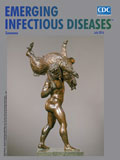
An Immortal Hero, an Enduring Challenge [PDF - 1.30 MB - 2 pages]
| EID | Breedlove B, Arguin PM. An Immortal Hero, an Enduring Challenge. Emerg Infect Dis. 2016;22(7):1328-1329. https://doi.org/10.3201/eid2207.ac2207 |
|---|---|
| AMA | Breedlove B, Arguin PM. An Immortal Hero, an Enduring Challenge. Emerging Infectious Diseases. 2016;22(7):1328-1329. doi:10.3201/eid2207.ac2207. |
| APA | Breedlove, B., & Arguin, P. M. (2016). An Immortal Hero, an Enduring Challenge. Emerging Infectious Diseases, 22(7), 1328-1329. https://doi.org/10.3201/eid2207.ac2207. |