Volume 23, Number 9—September 2017
Dispatch
Microcephaly Caused by Lymphocytic Choriomeningitis Virus
Maia Delaine , Anne-Sophie Weingertner, Antoine Nougairede, Quentin Lepiller, Samira Fafi-Kremer, Romain Favre, and Rémi N. Charrel
Figure
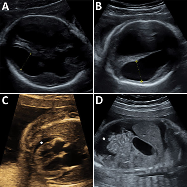
Figure. Ultrasonography of congenital microencephaly caused by infection with lymphocytic choriomeningitis virus diagnosed in the fetus of a 29-year-old pregnant women at 23 weeks’ gestation. A) Fetal brain at 23 weeks’ gestation showing symetric ventriculomegaly (14 mm). Yellow symbols indicate axis at which size of cerebral ventricle was measured. B) Fetal brain at 26 weeks’ gestation showing symetric ventriculomegaly (20 mm) and thinning of the cortical mantle. Yellow symbols indicate axis at which size of cerebral ventricle was measured. C) Fetal heart at 24 weeks’ gestation showing pericardial effusion (*) and cardiomyopathy with hyperechogenic muscle. D) Sagittal section of fetal abdomen at 26 weeks’ gestation showing ascites (*).
Page created: August 17, 2017
Page updated: August 17, 2017
Page reviewed: August 17, 2017
The conclusions, findings, and opinions expressed by authors contributing to this journal do not necessarily reflect the official position of the U.S. Department of Health and Human Services, the Public Health Service, the Centers for Disease Control and Prevention, or the authors' affiliated institutions. Use of trade names is for identification only and does not imply endorsement by any of the groups named above.