Volume 24, Number 5—May 2018
Dispatch
Transmission of Severe Fever with Thrombocytopenia Syndrome Virus by Haemaphysalis longicornis Ticks, China
Lu Zhuang1, Yi Sun1, Xiao-Ming Cui1, Fang Tang, Jian-Gong Hu, Li-Yuan Wang, Ning Cui, Zhen-Dong Yang, Dou-Dou Huang, Xiao-Ai Zhang, Wei Liu , and Wu-Chun Cao
Figure 2
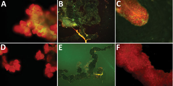
Figure 2. Specific detection of severe fever with thrombocytopenia syndrome virus (SFTSV) in tissues of adult Haemaphysalis longicornis ticks by indirect fluorescence assay. The green fluorescence represents the SFTS virus. A) Salivary gland of SFTSV-injected tick (original magnification ×40). B) Midgut of SFTSV-injected H. longicornis tick (original magnification ×10). C) Ovary of SFTSV-injected tick (original magnification ×40). D) Salivary gland of phosphate-buffered saline (PBS)–injected tick (original magnification ×40). E) Midgut of PBS-injected H. longicornis tick (original magnification ×10). F) Ovary of PBS-injected tick (original magnification ×40).
1These authors contributed equally to this article.
Page created: April 17, 2018
Page updated: April 17, 2018
Page reviewed: April 17, 2018
The conclusions, findings, and opinions expressed by authors contributing to this journal do not necessarily reflect the official position of the U.S. Department of Health and Human Services, the Public Health Service, the Centers for Disease Control and Prevention, or the authors' affiliated institutions. Use of trade names is for identification only and does not imply endorsement by any of the groups named above.