Human Usutu Virus Infection with Atypical Neurologic Presentation, Montpellier, France, 2016
Yannick Simonin

, Olivier Sillam, Marie J. Carles, Serafin Gutierrez, Patricia Gil, Orianne Constant, Marie F. Martin, Gilda Grard, Philippe Van de Perre, Sara Salinas, Isabelle Leparc-Goffart, and Vincent Foulongne
Author affiliations: Université de Montpellier, Montpellier, France (Y. Simonin, O. Constant, M.F. Martin, P. Van de Perre, S. Salinas, V. Foulongne); Université de Montpellier Hôpital, Montpellier (O. Sillam); Nimes University Hospital, Nimes, France (M.J. Carles); Centre de Coopération Internationale en Recherche Agronomique pour le Développement, Montpellier (S. Gutierrez, P. Gil); Institut de Recherche Biomédicale des Armées, Marseille, France (G. Grard, I. Leparc-Goffart)
Main Article
Figure 2
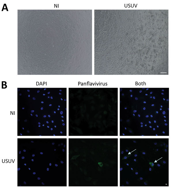
Figure 2. Cerebrospinal fluid sample of a 39-year-old man in Montpellier, France, infected with USUV who had an atypical neurologic presentation. The sample was amplified for 6 days on C6/36 cells, and the supernatant was used to infect Vero cells or primary human astrocytes. A) Cytopathic effect (presence of adherent dead cells and absence of heaps; all dead cells were scattered) was observed at day 5 postinfection of a Vero cell culture. Scale bar indicates 100 μm. B) Mock or infected primary human astrocytes were fixed at day 4 postinfection and labeled with pan-flavivirus antibody (MAB10216, clone D1–4G2) by indirect immunofluorescence (green). Strong labeling was observed in some cells (arrows). Nuclei are labeled with DAPI (4′,6-diamidino-2-phenylindole) (blue). NI, not infected; USUV, Usutu virus. Scale bar indicates 10 μm.
Main Article
Page created: April 24, 2018
Page updated: April 24, 2018
Page reviewed: April 24, 2018
The conclusions, findings, and opinions expressed by authors contributing to this journal do not necessarily reflect the official position of the U.S. Department of Health and Human Services, the Public Health Service, the Centers for Disease Control and Prevention, or the authors' affiliated institutions. Use of trade names is for identification only and does not imply endorsement by any of the groups named above.