Skip directly to site content Skip directly to page options Skip directly to A-Z link Skip directly to A-Z link Skip directly to A-Z link
Volume 25, Number 4—April 2019
Research Letter

Malignant Aspergillus flavus Otitis Externa with Jugular Thrombosis

Maxime Moniot, Marion Montava, Stéphane Ranque, Ugo Scemama, Carole CassagneComments to Author , and Varoquaux Arthur
Author affiliations: Aix-Marseille University, Marseille, France (M. Moniot, S. Ranque, C. Cassagne); La Conception University Hospital, Marseille (M. Montava, U. Scemama, V. Arthur)

Main Article

Figure

Magnetic resonance imaging (MRI) of a patient with malignant otitis externa, France. Cross-sectional imaging demonstrates a central skull base osteomyelitis in patient’s temporal bone. A) T1-weighted imaging; B, E) 3-dimensional T1-weighted imaging with gadolinium enhancement and fat saturation; C, F, G) T2-weighted imaging; and CT with iodine enhancement (D). Single asterisks (*) indicate jugular bulb thrombosis (panels B, D); double asterisks (**) indicate deep-spaces cellulitis (panels A–C).

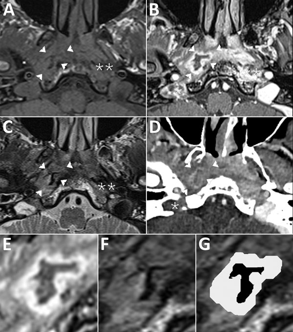
Figure. Magnetic resonance imaging (MRI) of a patient with malignant otitis externa, France. Cross-sectional imaging demonstrates a central skull base osteomyelitis in patient’s temporal bone. A) T1-weighted imaging; B, E) 3-dimensional T1-weighted imaging with gadolinium enhancement and fat saturation; C, F, G) T2-weighted imaging; and CT with iodine enhancement (D). Single asterisks (*) indicate jugular bulb thrombosis (panels B, D); double asterisks (**) indicate deep-spaces cellulitis (panels A–C). Arrowheads indicate parapharyngeal abscess at right (panels A–D); parapharyngeal abscess is also visible as a gray layer (panels E, G). The content of the abscess has an unusual “ink smudge” pattern with no signal in T2-weighted imaging, visible as a black layer (panels F, G). This pattern is consistent with a mycetoma surrounded by granulation tissue.

Main Article

Page created: March 17, 2019
Page updated: March 17, 2019
Page reviewed: March 17, 2019
The conclusions, findings, and opinions expressed by authors contributing to this journal do not necessarily reflect the official position of the U.S. Department of Health and Human Services, the Public Health Service, the Centers for Disease Control and Prevention, or the authors' affiliated institutions. Use of trade names is for identification only and does not imply endorsement by any of the groups named above.
file_external