Volume 26, Number 2—February 2020
Research Letter
Human Alveolar Echinococcosis, Croatia
Davorka Dušek, Adriana Vince, Ivan Kurelac, Neven Papić, Klaudija Višković, Peter Deplazes, and Relja Beck
Figure
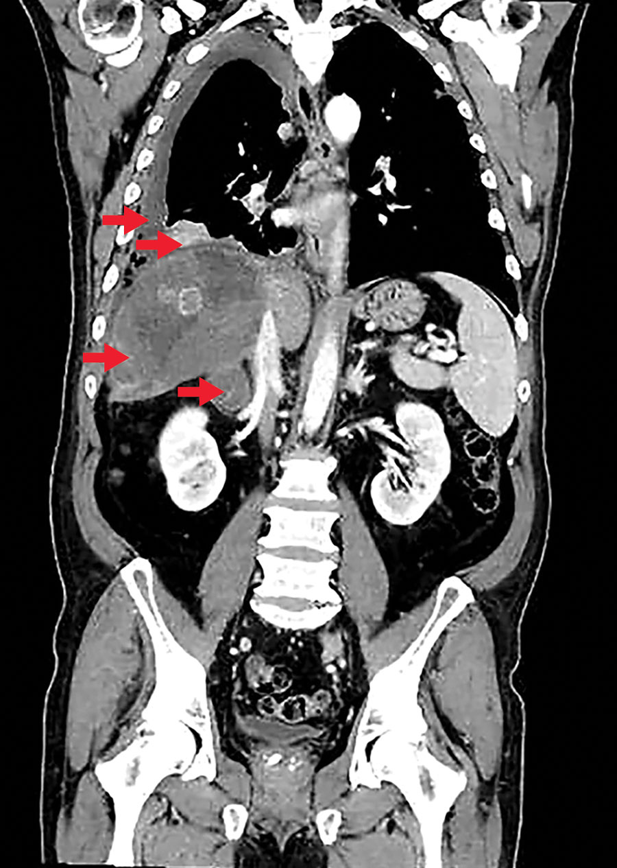
Figure. Computed tomography scan of a patient diagnosed with alveolar echinococcosis, Croatia. Arrows indicate right pleural effusion, lung lesions, an enlarged right adrenal gland, and a 13 × 12 × 12 cm lesion in the liver caused by Echinococcus multilocaris.
Page created: January 20, 2020
Page updated: January 20, 2020
Page reviewed: January 20, 2020
The conclusions, findings, and opinions expressed by authors contributing to this journal do not necessarily reflect the official position of the U.S. Department of Health and Human Services, the Public Health Service, the Centers for Disease Control and Prevention, or the authors' affiliated institutions. Use of trade names is for identification only and does not imply endorsement by any of the groups named above.