Skip directly to site content Skip directly to page options Skip directly to A-Z link Skip directly to A-Z link Skip directly to A-Z link
Volume 26, Number 2—February 2020
Dispatch

Ocular Spiroplasma ixodetis in Newborns, France

Alexandre MatetComments to Author , Anne Le Flèche-Matéos, François Doz, Pascal Dureau, and Nathalie Cassoux
Author affiliations: Institut Curie and Université Paris Descartes, Paris, France (A. Matet, F. Doz, N. Cassoux); Institut Pasteur, ​Paris (A. Le Flèche-Matéos); Fondation Ophtalmologique Adolphe de Rothschild, Paris (P. Dureau)

Main Article

Figure 1

Ocular anterior segment in 3 newborn infants with bilateral total cataract and anterior uveitis related to endogenous Spiroplasma ixodetis infection. A, B) Case-patient 1. Right (A) and left (B) eyes of a 4-week-old girl showing total cataract, posterior synechiae due to a cyclitic fibrinic membrane, and large keratic precipitates more visible in the left eye. The immature iris vasculature is dilated in the context of anterior segment inflammation. C, D) Case-patient 2. Right (C) and left (D) ey

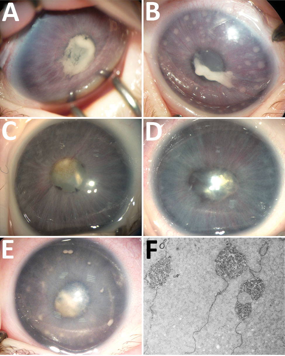
Figure 1. Ocular anterior segment in 3 newborn infants with bilateral total cataract and anterior uveitis related to endogenous Spiroplasma ixodetis infection. A, B) Case-patient 1. Right (A) and left (B) eyes of a 4-week-old girl showing total cataract, posterior synechiae due to a cyclitic fibrinic membrane, and large keratic precipitates more visible in the left eye. The immature iris vasculature is dilated in the context of anterior segment inflammation. C, D) Case-patient 2. Right (C) and left (D) eyes of a 6-week-old boy showing total cataract, posterior synechiae, dilated immature iris vessels, and few keratic precipitates more visible in the left eye. E) Case-patient 3. Left eye of a 1-month-old boy with multiple retrocorneal white deposits, total cataract, posterior synechiae, and immature dilated iris vessels. F) Case-patient 3. Electron transmission microscopy of crystalline lens material from a 2-month-old boy with total cataract and anterior uveitis, revealing the presence of microorganisms with spiral-like projections highly suggestive of bacteria from the Spiroplasma genus.

Main Article

Page created: January 20, 2020
Page updated: January 20, 2020
Page reviewed: January 20, 2020
The conclusions, findings, and opinions expressed by authors contributing to this journal do not necessarily reflect the official position of the U.S. Department of Health and Human Services, the Public Health Service, the Centers for Disease Control and Prevention, or the authors' affiliated institutions. Use of trade names is for identification only and does not imply endorsement by any of the groups named above.
file_external