Skip directly to site content Skip directly to page options Skip directly to A-Z link Skip directly to A-Z link Skip directly to A-Z link
Volume 26, Number 9—September 2020
Synopsis

Pathology and Pathogenesis of SARS-CoV-2 Associated with Fatal Coronavirus Disease, United States

Roosecelis B. Martines1Comments to Author , Jana M. Ritter1, Eduard Matkovic, Joy Gary, Brigid C. Bollweg, Hannah Bullock, Cynthia S. Goldsmith, Luciana Silva-Flannery, Josilene N. Seixas, Sarah Reagan-Steiner, Timothy Uyeki, Amy Denison, Julu Bhatnagar, Wun-Ju Shieh, Sherif R. Zaki, and COVID-19 Pathology Working Group
Author affiliations: Centers for Disease Control and Prevention, Atlanta, Georgia, USA (R.B. Martines, J.M. Ritter, E. Matkovic, J. Gary, B.C. Bollweg, C.S. Goldsmith, L. Silva-Flannery, J.N. Seixas, S. Reagan-Steiner, T. Uyeki, A. Denison, J. Bhatnagar, W.-J. Shieh, S.R. Zaki); Synergy America Inc., Atlanta (H. Bullock).

Main Article

Figure 1

Pulmonary histopathology in fatal coronavirus disease cases caused by severe acute respiratory syndrome coronavirus 2 infection. A) Patient no. 5: tracheitis characterized by moderate mononuclear inflammation within the submucosa (original magnification ×10). B) Patient no. 3: extensive denudation of tracheal epithelium; submucosal congestion, mild edema, and mononuclear inflammation (original magnification ×10). C) Patient no. 4: exudative phase of diffuse alveolar damage characterized by abund

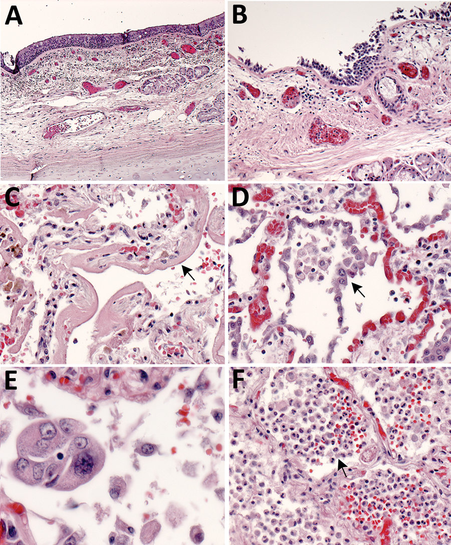
Figure 1. Pulmonary histopathology in fatal coronavirus disease cases caused by severe acute respiratory syndrome coronavirus 2 infection. A) Patient no. 5: tracheitis characterized by moderate mononuclear inflammation within the submucosa (original magnification ×10). B) Patient no. 3: extensive denudation of tracheal epithelium; submucosal congestion, mild edema, and mononuclear inflammation (original magnification ×10). C) Patient no. 4: exudative phase of diffuse alveolar damage characterized by abundant hyaline membranes lining alveolar spaces (arrow) (original magnification ×20). D) Patient no. 8: proliferative phase of diffuse alveolar damage characterized by proliferation of type II pneumocytes (arrow) (original magnification ×20). E) Patient no. 1: atypical pneumocytes with enlarged and multiple nuclei, and expanded cytoplasm in a case with proliferative DAD (original magnification ×40). F) Patient no. 7: bronchopneumonia with filling of alveolar spaces by neutrophils and patchy hemorrhage (arrow) (original magnification ×10).

Main Article

1These authors contributed equally to this article.

2Members of the COVID-19 Pathology Working Group: Rhonda Cole, Amanda Lewis, Pamela Fair, Lindsey Estetter.

Page created: May 21, 2020
Page updated: August 18, 2020
Page reviewed: August 18, 2020
The conclusions, findings, and opinions expressed by authors contributing to this journal do not necessarily reflect the official position of the U.S. Department of Health and Human Services, the Public Health Service, the Centers for Disease Control and Prevention, or the authors' affiliated institutions. Use of trade names is for identification only and does not imply endorsement by any of the groups named above.
file_external