Skip directly to site content Skip directly to page options Skip directly to A-Z link Skip directly to A-Z link Skip directly to A-Z link
Volume 26, Number 9—September 2020
Synopsis

Pathology and Pathogenesis of SARS-CoV-2 Associated with Fatal Coronavirus Disease, United States

Roosecelis B. Martines1Comments to Author , Jana M. Ritter1, Eduard Matkovic, Joy Gary, Brigid C. Bollweg, Hannah Bullock, Cynthia S. Goldsmith, Luciana Silva-Flannery, Josilene N. Seixas, Sarah Reagan-Steiner, Timothy Uyeki, Amy Denison, Julu Bhatnagar, Wun-Ju Shieh, Sherif R. Zaki, and COVID-19 Pathology Working Group
Author affiliations: Centers for Disease Control and Prevention, Atlanta, Georgia, USA (R.B. Martines, J.M. Ritter, E. Matkovic, J. Gary, B.C. Bollweg, C.S. Goldsmith, L. Silva-Flannery, J.N. Seixas, S. Reagan-Steiner, T. Uyeki, A. Denison, J. Bhatnagar, W.-J. Shieh, S.R. Zaki); Synergy America Inc., Atlanta (H. Bullock).

Main Article

Figure 5

Ultrastructural features of severe acute respiratory syndrome coronavirus 2 infection within the upper airway of a fatal coronavirus disease case from formalin-fixed paraffin-embedded (FFPE) tissue. Viral particles associated with the cilia of ciliated cells (A, C, and D) and the cytoplasm of respiratory epithelial cells (B) in the upper airway are indicted by arrows. Images in panels A and C were obtained from FFPE tissue removed from a paraffin block using a 2-mm biopsy punch. Images in panels

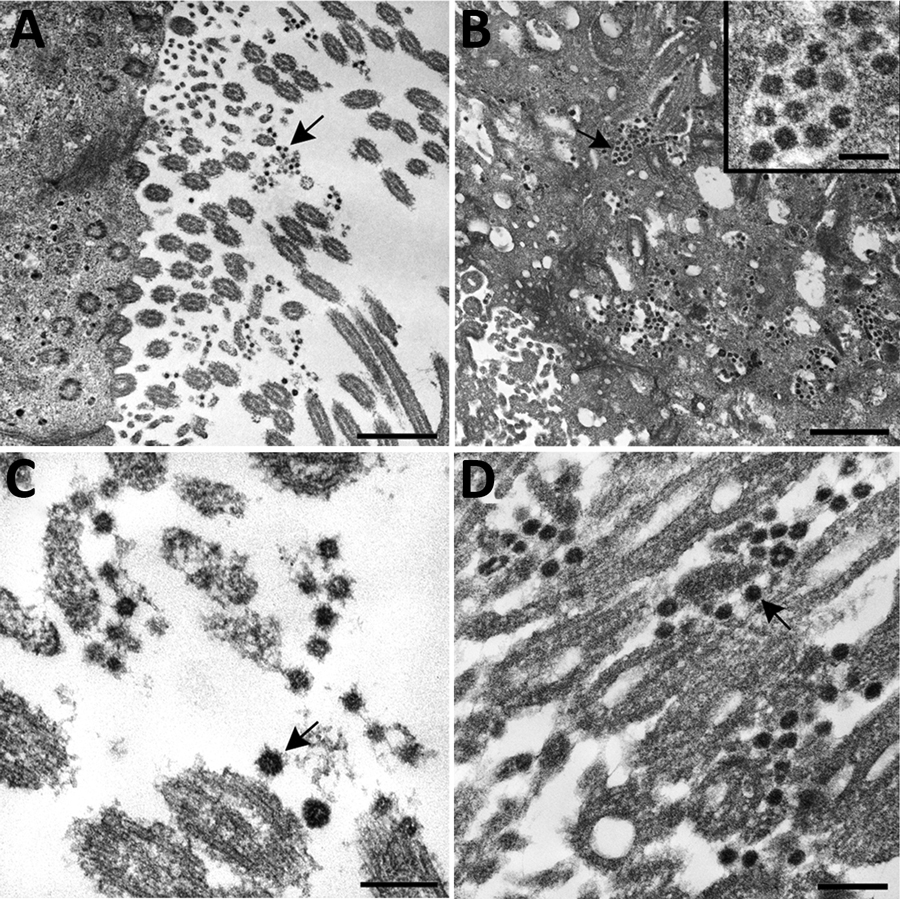
Figure 5. Ultrastructural features of severe acute respiratory syndrome coronavirus 2 infection within the upper airway of a fatal coronavirus disease case from formalin-fixed paraffin-embedded (FFPE) tissue. Viral particles associated with the cilia of ciliated cells (A, C, and D) and the cytoplasm of respiratory epithelial cells (B) in the upper airway are indicted by arrows. Images in panels A and C were obtained from FFPE tissue removed from a paraffin block using a 2-mm biopsy punch. Images in panels B and D were collected from a 3 μm section of FFPE tissue affixed to a glass slide. Viral particles visualized in FFPE samples were smaller than those observed from fresh tissue; extracellular viral particles in fresh tissue samples were 105 nm in diameter and those from FFPE tissues were 75 nm in diameter. Scale bars indicate 1 μm (panel A), 800 nm (panel B), and 200 nm (panels C and D).

Main Article

1These authors contributed equally to this article.

2Members of the COVID-19 Pathology Working Group: Rhonda Cole, Amanda Lewis, Pamela Fair, Lindsey Estetter.

Page created: May 21, 2020
Page updated: August 18, 2020
Page reviewed: August 18, 2020
The conclusions, findings, and opinions expressed by authors contributing to this journal do not necessarily reflect the official position of the U.S. Department of Health and Human Services, the Public Health Service, the Centers for Disease Control and Prevention, or the authors' affiliated institutions. Use of trade names is for identification only and does not imply endorsement by any of the groups named above.
file_external