Volume 27, Number 12—December 2021
Research
Novel Filoviruses, Hantavirus, and Rhabdovirus in Freshwater Fish, Switzerland, 2017
Melanie M. Hierweger, Michel C. Koch, Melanie Rupp, Piet Maes, Nicholas Di Paola, Rémy Bruggmann, Jens H. Kuhn, Heike Schmidt-Posthaus1, and Torsten Seuberlich1
Figure 4
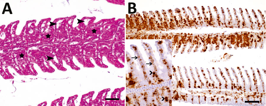
Figure 4. Histopathologic lesions and viral RNA in European perch infected with Bern perch virus. A) Histopathologic lesions in gills (hematoxylin and eosin stain) showing epithelial hypertrophy and hyperplasia, multifocally leading to lamellar fusion (stars) and multifocal epithelial lifting due to edema (closed arrowheads). Scale bar indicates 25 μm. B) In situ hybridization detection of Bern perch virus RNA in gills (brown labeling): positive macrophages, more pronounced in proliferated areas, and endothelial cells. Inset: higher magnification showing positive macrophages (open arrowheads) and endothelial cells (arrows with open heads). Scale bar indicates 50 μm.
1These authors contributed equally to this article.
Page created: October 06, 2021
Page updated: November 22, 2021
Page reviewed: November 22, 2021
The conclusions, findings, and opinions expressed by authors contributing to this journal do not necessarily reflect the official position of the U.S. Department of Health and Human Services, the Public Health Service, the Centers for Disease Control and Prevention, or the authors' affiliated institutions. Use of trade names is for identification only and does not imply endorsement by any of the groups named above.