Skip directly to site content Skip directly to page options Skip directly to A-Z link Skip directly to A-Z link Skip directly to A-Z link
Volume 27, Number 9—September 2021
Etymologia

Talaromyces marneffei

Monika MahajanComments to Author 
Author affiliation: Postgraduate Institute of Medical Education and Research, Chandigarh, India

Main Article

Figure 2

A) Ultrastructural morphology of Talaromyces marneffei, including chains of single-celled, teardrop-shaped conidia, each originating from its respective, flask-shaped phialide. Source: Libero Ajello, Centers for Disease Control and Prevention (https://phil.cdc.gov/Details.aspx?pid = 4240). B) Superior (front) view of a petri dish culture plate on which a wrinkled colony of Penicillium marneffei has been cultivated. Source: James Gathany, Centers for Disease Control and Prevention (https://phil.cdc.gov/Details.aspx?pid = 1879). C) Mouse testicle tissue specimen showing globe-shaped yeast cells of T. marneffei undergoing multiplication by binary fission not by mitosis (methenamine silver stain). Source: Libero Ajello, Centers for Disease Control and Prevention (https://phil.cdc.gov/Details.aspx?pid = 4235); D) Gradual conversion of mycelial phase of T. marneffei (growth at 25°C) to yeast phase on brain heart infusion agar after incubation at 37°C. Mycelial phase (first tube marked 25°C) shows diffusible red pigment. Source: Monika Mahajan, Postgraduate Institute of Medical Education and Research, Chandigarh, India; E) Loose network of hyphae of T. marneffei forming gymnothecium that contains asci. Source: https://istudy.pk/ascomycota-fruit-bodies/.

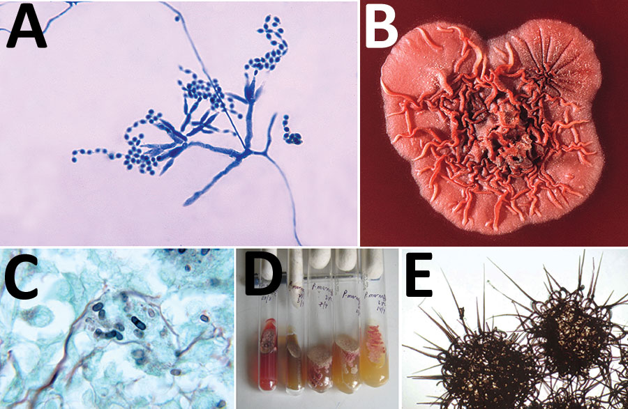
Figure 2. A) Ultrastructural morphology of Talaromyces marneffei, including chains of single-celled, teardrop-shaped conidia, each originating from its respective, flask-shaped phialide. Source: Libero Ajello, Centers for Disease Control and Prevention (https://phil.cdc.gov/Details.aspx?pid = 4240). B) Superior (front) view of a petri dish culture plate on which a wrinkled colony of Penicillium marneffei has been cultivated. Source: James Gathany, Centers for Disease Control and Prevention (https://phil.cdc.gov/Details.aspx?pid = 1879). C) Mouse testicle tissue specimen showing globe-shaped yeast cells of T. marneffei undergoing multiplication by binary fission not by mitosis (methenamine silver stain). Source: Libero Ajello, Centers for Disease Control and Prevention (https://phil.cdc.gov/Details.aspx?pid = 4235); D) Gradual conversion of mycelial phase of T. marneffei (growth at 25°C) to yeast phase on brain heart infusion agar after incubation at 37°C. Mycelial phase (first tube marked 25°C) shows diffusible red pigment. Source: Monika Mahajan, Postgraduate Institute of Medical Education and Research, Chandigarh, India; E) Loose network of hyphae of T. marneffei forming gymnothecium that contains asci. Source: https://istudy.pk/ascomycota-fruit-bodies/.

Main Article

Page created: August 09, 2021
Page updated: August 17, 2021
Page reviewed: August 17, 2021
The conclusions, findings, and opinions expressed by authors contributing to this journal do not necessarily reflect the official position of the U.S. Department of Health and Human Services, the Public Health Service, the Centers for Disease Control and Prevention, or the authors' affiliated institutions. Use of trade names is for identification only and does not imply endorsement by any of the groups named above.
file_external