Volume 27, Number 9—September 2021
Etymologia
Talaromyces marneffei
Talaromyces marneffei [t læ′ ɹɒ maɪ̯s ɪz mɑ:neɪ′]
Figure 1

Figure 1. Hubert Marneffe (1901‒1970) Source: Wikimanche, Institut Pasteur, public domain.
Talaromyces marneffei (formerly Penicillium marneffei) is a thermally dimorphic fungus that causes talaromycosis, which was previously called penicilliosis. The genus name Talaromyces is derived from the Greek words tálaros (basket) and múkēs (mushroom). Talaros aptly describes the ascocarp known as a gymnothecium (composed of fine woven hyphae) in which asci are formed. Asexual stages of Talaromyces species were previously known as the species Penicillium of the subgenus Biverticillium. Capponi and Sureau isolated the fungus at Institute Pasteur de Dalat in Vietnam in 1955 from Chinese bamboo rats (Rhizomys sinensis). In 1959, Gabriel Segretain, after an accidental finger prick with a needle containing the yeast cells, described the fungus as a new species, naming it Penicillium marneffei in honor of Hubert Marneffe (1901‒1970), the Director of the Institute in Indochina (Figure 1).
Figure 2
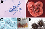
Figure 2. A) Ultrastructural morphology of Talaromyces marneffei, including chains of single-celled, teardrop-shaped conidia, each originating from its respective, flask-shaped phialide. Source: Libero Ajello, Centers for Disease Control and Prevention...
Talaromycosis affects persons who live in or visit Southeast Asia, southern China, or northeastern India, and are immunocompromised because of HIV/AIDS, cancer, organ transplant, or adult-onset immunodeficiency syndrome (Figure 2). This disease occurs after inhalation of aerosolized fungal spores from the environment. Although the precise reservoir is unknown, T. marneffei is found in bamboo rats.
References
- Pitt JI. Penicillium and Talaromyces. In: Batt C. Patel P, editors. Encyclopedia of food microbiology. New York: Elsevier; 2014. p. 6–13.
- Talaromycosis (formerly penicilliosis) [cited 2021 Jun 10]. https://www.cdc.gov/fungal/diseases/other/talaromycosis.html
- Tsang C-C, Lau SKP, Woo PCY. Sixty years from Segretain’s description: what have we learned and should learn about the basic mycology of Talaromyces marneffei? Mycopathologia. 2019;184:721–9. DOIPubMedGoogle Scholar
- Vanittanakom N, Cooper CR Jr, Fisher MC, Sirisanthana T. Penicillium marneffei infection and recent advances in the epidemiology and molecular biology aspects. Clin Microbiol Rev. 2006;19:95–110. DOIPubMedGoogle Scholar
Figures
Cite This ArticleOriginal Publication Date: August 09, 2021
Related Links
Table of Contents – Volume 27, Number 9—September 2021
| EID Search Options |
|---|
|
|
|
|
|
|
Please use the form below to submit correspondence to the authors or contact them at the following address:
Monika Mahajan, Postgraduate Institute of Medical Education and Research, Medical Microbiology, Research Block A, Sector 12, Chandigarh 160012, India
Top