Volume 29, Number 1—January 2023
Dispatch
Clinical Forms of Japanese Spotted Fever from Case-Series Study, Zigui County, Hubei Province, China, 2021
Abstract
We report a case-series study of 5 patients with Japanese spotted fever from the Three Gorges Area in China, including 1 fatal case. Seroprevalence of Rickettsia japonica was ≈21% among the local population. Our report highlights the emerging potential threat to human health of Japanese spotted fever in the area.
Japanese spotted fever (JSF), caused by the bacterium Rickettsia japonica, was first described in 1984 in Japan (1). It has been recognized in multiple countries in Asia, including Japan, South Korea, the Philippines, Thailand, and China (1–5), suggesting that it is endemic in Asia. In China, R. japonica has been detected in ticks from the central, southeast, and northeast regions (6–8). Since JSF cases were first found in Anhui Province in 2013 (5), a total of 39 have been reported in the Dabie Mountains on the borders of Henan, Anhui, and Hubei Provinces and in the Tianmu Mountains in Zhejiang Province on the eastern coast (Appendix Figure 1) (9–11).
Our study was approved by the ethics committees of the National Institute for Communicable Disease Control and Prevention (ICDC). During April–October 2021, five JSF cases were found in Zigui County, Hubei Province, in the Three Gorges area of China (Appendix Figure 1), including case 1, in which the patient died because of multiorgan failure. Among the 5 case-patients (3 female, 2 male), disease onset occurred during April–October (2 cases in spring, 3 in autumn); median age of case-patients was 53 years (range 47–70 years) (Table 1). All had histories of activity in fields, but none was aware of any tick bites. All initially manifested signs and symptoms of sudden fever (≥38.5°C), headache, chills, fatigue, chest tightness, shortness of breath, and dyspnea. All had rash and eschar (Appendix Figure 2), except for case-patient 1, who had no eschar. Case-patients 1, 3, and 5 experienced vomiting and case-patient 3 had abdominal pain. Medical laboratory tests (Table 2) showed that case-patients 1, 3, and 5 had thrombocytopenia and lymphopenia, case-patients 1 and 5 had neutrophilia, and case-patients 1 and 4 had mild anemia. All 5 patients had remarkably elevated levels of C-reactive protein and D-dimer, and except for case-patient 5, all had elevated liver-associated enzymes. Proteinuria was detected in case-patients 1, 2, 3, and 5. Case-patients 1, 3, and 5 had simultaneous edema and effusion and case-patient 5 showed multiple effusions (pelvic, pleural, pericardial, and ascites).
Most laboratory tests were flagged as abnormal for case-patient 1, who died. She received an initial antimicrobial treatment (Appendix) at the village clinic without a causative agent being identified and already had severe sepsis (sequential organ failure assessment score = 12) by the time she was admitted to the hospital on day 8 after symptom onset. Disseminated intravascular coagulation was diagnosed, and she died of multiple organ failure on day 9 despite continuous intensive therapy. The other 4 patients recovered without sequelae after receiving doxycycline or minocycline treatment (Appendix).
Figure
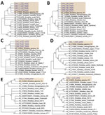
Figure. Bootstrap consensus phylogenetic tree constructed based on partial sequences of 17kDa (A), gltA (B), groEL (C), ompA (D), ompB (E), and ...
Using a PCR assay, we detected 6 rickettsial genes (groEL, gltA, ompA, ompB, sca4, and 17kDa gene) in a blood specimen from case-patient 1. In addition, we amplified groEL, gltA, and 17kDa genes from case-patients 2, 3, and 5 but only gltA and 17kDa from case-patient 4. For all 5 patients, the genomic sequences of each amplified target gene were 100% identical to each other; phylogenetic analysis revealed that the causative agent was most closely related to R. japonica (Figure). We submitted the obtained sequences to GenBank (accession nos. OM966424–8 for gltA, OM966422 for ompA, OM966423 for ompB, OM966412–6 for 17kDa, OM966417 for sca4, and OM966418–21 for groEL). We obtained 1 stable rickettsial isolate, designated R. japonica strain YC21, from the blood of case-patient 1 using Vero cell cultures (Appendix Figure 3) and obtained the whole genomic sequence from the isolate (GenBank BioProject PRJNA812951). Phylogenetic analysis based on core genes suggested that R. japonica strain YC21 was most closely related to R. japonica strain LA16/2015 (Appendix Figure 4); 30 virulence-associated genes of R. japonica strain YC21 (Appendix Table 2), predicted using the virulence factor database (http://www.mgc.ac.cn/VFs), were completely homologous to those of strain LA16/2015.
We used an immunofluorescence assay using R. japonica strain YC21 as coating antigen (a cutoff of 1:64 was determined by testing negative and positive samples) and R. rickettsii (Focus Diagnostics, http://focusdiagnostics.in) to test serum-specific antibodies from the 5 JSF patients and 100 healthy subjects recruited locally. Case-patients 2 and 3 were confirmed to have JSF on the basis of a ≥4-fold increase in R. rickettsii–specific and R. japonica–specific IgG titers between acute and convalescent phase serum (Appendix Table 3). At baseline, 12/100 (12%) of the healthy local donors tested positive (range, 1:128–1:1,024; geometric mean, 512) for R. japonica–specific IgG.
We measured cytokine and chemokine levels in the serum samples collected from the JSF patients (during acute phase) and 6 healthy donors (Appendix Table 4). The levels of interferon (IFN) γ, interleukin (IL) 6, IL-10, IL-1α, macrophage inflammatory protein 1β, IL-8, IFN gamma-induced protein 10, and monocyte chemoattractant protein 1 in the 4 surviving JSF patients were significantly higher than in the healthy donors (p <0.01), consistent with previous reports, except for the exclusion of tumor necrosis factor α (12–14). In the case-patient who died, serum levels of IL-6, IL-10, and IFN-γ were 10-fold higher than those in the surviving case-patients and the levels of IL-4, INF-α, granulocyte-macrophage colony-stimulating factor, monocyte chemoattractant protein 1, macrophage inflammatory protein 1β, and IP-10 were 2-fold higher, suggesting that R. japonica infection might cause an unregulated hyperinflammatory state, potentially leading to cytokine release syndrome (14).
We identified 5 cases of JSF, including 1 in which the patient died, in Zigui County in the Three Gorges Area of China, where JSF has not previously been identified. Furthermore, our study revealed a high prevalence (12%) of R. japonica among residents, suggesting a new endemic area for JSF in China and indicating that JSF might be more widespread than previously thought. We should be alert to the potential risk for JSF, especially in areas where R. japonica is detected in vectors (Appendix Figure 1). The JSF cases were confirmed by PCR detection and serologic tests. A strain of R. japonica isolated from the blood of the patient who died was revealed to be most closely related to strains LA16/2015 and LA4/2015 detected in Zhejiang Province, suggesting that a virulent strain of R. japonica might have spread widely across China.
Delayed treatment is one of the worst prognostic factors for patients with JSF, and as a neglected infectious disease, it might not be considered during differential diagnosis. In our study, the patient who died manifested a faint rash, but without eschar, which resulted in delayed diagnosis and provision of correct antimicrobial treatment when she first visited the rural clinic. Profiling the patient’s serum cytokine and chemokine levels indicated notably elevated IL-6, IL-10, and IFN-γ, characteristic of potential cytokine release syndrome. The primary findings on patient cytokines levels benefit understanding of immune response to R. japonica infection.
Our findings highlight the threat of JSF to public health in China. Healthcare workers, especially in rural areas where residents are at increased risk for tick exposure, should be aware of this potentially deadly infectious disease. Long-term surveillance and investigation of local hosts and vectors of R. japonica are necessary to improve the prevention and treatment of JSF.
Dr. Teng is a research associate at the National Institute for Communicable Disease Control and Prevention, Chinese Center for Disease Control and Prevention. His research interests are detection and isolation of rickettsia and the epidemiology of rickettsioses.
Acknowledgments
We thank Jingdong Song for his assistance with acquiring scanning electron microscope images.
This work was supported by grants from the National Natural Science Foundation of China (grant no. 81671985), the Science Foundation for the State Key Laboratory for Infectious Disease Prevention and Control from China (grant no. 2019SKLID403, 2021SKLID507), and the Pathogen Monitoring Capability Improvement Project (grant no. 131031102000150003) from the National Health Commission of China.
References
- Mahara F. Japanese spotted fever: report of 31 cases and review of the literature. Emerg Infect Dis. 1997;3:105–11. DOIPubMedGoogle Scholar
- Camer GA, Alejandria M, Amor M, Satoh H, Muramatsu Y, Ueno H, et al. Detection of antibodies against spotted fever group Rickettsia (SFGR), typhus group Rickettsia (TGR), and Coxiella burnetii in human febrile patients in the Philippines. Jpn J Infect Dis. 2003;56:26–8.PubMedGoogle Scholar
- Chung MH, Lee SH, Kim MJ, Lee JH, Kim ES, Lee JS, et al. Japanese spotted fever, South Korea. Emerg Infect Dis. 2006;12:1122–4. DOIPubMedGoogle Scholar
- Gaywee J, Sunyakumthorn P, Rodkvamtook W, Ruang-areerate T, Mason CJ, Sirisopana N. Human infection with Rickettsia sp. related to R. japonica, Thailand. Emerg Infect Dis. 2007;13:657–9. DOIPubMedGoogle Scholar
- Li J, Hu W, Wu T, Li HB, Hu W, Sun Y, et al. Japanese spotted fever in eastern China, 2013. Emerg Infect Dis. 2018;24:2107–9. DOIPubMedGoogle Scholar
- Zhang L, Cui F, Wang L, Zhang L, Zhang J, Wang S, et al. Investigation of anaplasmosis in Yiyuan County, Shandong Province, China. Asian Pac J Trop Med. 2011;4:568–72. DOIPubMedGoogle Scholar
- Yan P, Qiu Z, Zhang T, Li Y, Wang W, Li M, et al. Microbial diversity in the tick Argas japonicus (Acari: Argasidae) with a focus on Rickettsia pathogens. Med Vet Entomol. 2019;33:327–35. DOIPubMedGoogle Scholar
- Lu M, Li F, Liao Y, Shen JJ, Xu JM, Chen YZ, et al. Epidemiology and diversity of Rickettsiales bacteria in humans and animals in Jiangsu and Jiangxi Provinces, China. Sci Rep. 2019;9:13176. DOIPubMedGoogle Scholar
- Lu Q, Yu J, Yu L, Zhang Y, Chen Y, Lin M, et al. Rickettsia japonica infections in humans, Zhejiang Province, China, 2015. Emerg Infect Dis. 2018;24:2077–9. DOIPubMedGoogle Scholar
- Li W, Liu SN. Rickettsia japonica infections in Huanggang, China, in 2021. IDCases. 2021;26:
e01309 . DOIPubMedGoogle Scholar - Li H, Zhang PH, Du J, Yang ZD, Cui N, Xing B, et al. Rickettsia japonica infections in humans, Xinyang, China, 2014–2017. Emerg Infect Dis. 2019;25:1719–22. DOIPubMedGoogle Scholar
- Popivanova NI, Murdjeva MA, Baltadzhiev IG, Haydushka IA. Dynamics in serum cytokine responses during acute and convalescent stages of Mediterranean spotted fever. Folia Med (Plovdiv). 2011;53:36–43. DOIPubMedGoogle Scholar
- Kondo M, Matsushima Y, Mizutani K, Iida S, Habe K, Yamanaka K. Transition of serum cytokine concentration in Rickettsia japonica infection. Infect Dis Rep. 2020;12:127–31. DOIPubMedGoogle Scholar
- Tai K, Iwasaki H, Ikegaya S, Takada N, Tamaki Y, Tabara K, et al. Significantly higher cytokine and chemokine levels in patients with Japanese spotted fever than in those with Tsutsugamushi disease. J Clin Microbiol. 2014;52:1938–46. DOIPubMedGoogle Scholar
Figure
Tables
Cite This Article1These authors contributed equally to this article.
Table of Contents – Volume 29, Number 1—January 2023
| EID Search Options |
|---|
|
|
|
|
|
|
Please use the form below to submit correspondence to the authors or contact them at the following address:
Tian Qin, State Key Laboratory of Infectious Diseases Prevention and Control, National Institute for Communicable Disease Control and Prevention, Chinese Center for Disease Control and Prevention, 155 Changbai Rd, Changping, Beijing 102206, China
Top