Synopses
The emergence of SARS-CoV-2 and the worldwide COVID-19 pandemic triggered considerable attention to the emergence and evolution of novel human pathogens. Bourbon virus (BRBV) was first discovered in 2014 in Bourbon County, Kansas, USA. Since its initial discovery, several cases of BRBV infection in humans have been identified in Kansas, Oklahoma, and Missouri. BRBV is classified within the Thogotovirus genus; these negative-strand RNA viruses appear to be transmitted by ticks, and much of their biology remains unknown. In this review, we describe the emergence, virology, geographic range and ecology, and human disease caused by BRBV and discuss potential treatments for active BRBV infections. This virus and other emerging viral pathogens remain key public health concerns and require continued surveillance and study to mitigate human exposure and disease.
| EID | Roe MK, Huffman ER, Batista YS, Papadeas GG, Kastelitz SR, Restivo AM, et al. Comprehensive Review of Emergence and Virology of Tickborne Bourbon Virus in the United States. Emerg Infect Dis. 2023;29(1):1-7. https://doi.org/10.3201/eid2901.212295 |
|---|---|
| AMA | Roe MK, Huffman ER, Batista YS, et al. Comprehensive Review of Emergence and Virology of Tickborne Bourbon Virus in the United States. Emerging Infectious Diseases. 2023;29(1):1-7. doi:10.3201/eid2901.212295. |
| APA | Roe, M. K., Huffman, E. R., Batista, Y. S., Papadeas, G. G., Kastelitz, S. R., Restivo, A. M....Stobart, C. C. (2023). Comprehensive Review of Emergence and Virology of Tickborne Bourbon Virus in the United States. Emerging Infectious Diseases, 29(1), 1-7. https://doi.org/10.3201/eid2901.212295. |
We performed a case–control study across 25 hospitals in India for the period of January–June 2021 to evaluate the reasons for an COVID-19–associated mucormycosis (CAM) outbreak. We investigated whether COVID-19 treatment practices (glucocorticoids, zinc, tocilizumab, and others) were associated with CAM. We included 1,733 cases of CAM and 3,911 age-matched COVID-19 controls. We found cumulative glucocorticoid dose (odds ratio [OR] 1.006, 95% CI 1.004–1.007) and zinc supplementation (OR 2.76, 95% CI 2.24–3.40), along with elevated C-reactive protein (OR 1.004, 95% CI 1.002–1.006), host factors (renal transplantation [OR 7.58, 95% CI 3.31–17.40], diabetes mellitus [OR 6.72, 95% CI 5.45–8.28], diabetic ketoacidosis during COVID-19 [OR 4.41, 95% CI 2.03–9.60]), and rural residence (OR 2.88, 95% CI 2.12–3.79), significantly associated with CAM. Mortality rate at 12 weeks was 32.2% (473/1,471). We emphasize the judicious use of COVID-19 therapies and optimal glycemic control to prevent CAM.
| EID | Muthu V, Agarwal R, Rudramurthy S, Thangaraju D, Shevkani M, Patel AK, et al. Multicenter Case–Control Study of COVID-19–Associated Mucormycosis Outbreak, India. Emerg Infect Dis. 2023;29(1):8-19. https://doi.org/10.3201/eid2901.220926 |
|---|---|
| AMA | Muthu V, Agarwal R, Rudramurthy S, et al. Multicenter Case–Control Study of COVID-19–Associated Mucormycosis Outbreak, India. Emerging Infectious Diseases. 2023;29(1):8-19. doi:10.3201/eid2901.220926. |
| APA | Muthu, V., Agarwal, R., Rudramurthy, S., Thangaraju, D., Shevkani, M., Patel, A. K....Chakrabarti, A. (2023). Multicenter Case–Control Study of COVID-19–Associated Mucormycosis Outbreak, India. Emerging Infectious Diseases, 29(1), 8-19. https://doi.org/10.3201/eid2901.220926. |
Role of Seaports and Imported Rats in Seoul Hantavirus Circulation, Africa [PDF - 591 KB - 6 pages]
Seoul orthohantavirus (SEOV) is not considered a major public health threat on the continent of Africa. However, Africa is exposed to rodentborne SEOV introduction events through maritime traffic after exponential growth of trade with the rest of the world. Serologic studies have already detected hantavirus antibodies in human populations, and recent investigations have confirmed circulation of hantavirus, including SEOV, in rat populations. Thus, SEOV is a possible emerging zoonotic risk in Africa. Moreover, the range of SEOV could rapidly expand, and transmission to humans could increase because of host switching from the usual brown rat (Rattus norvegicus) species, which is currently invading Africa, to the more widely installed black rat (R. rattus) species. Because of rapid economic development, environmental and climatic changes, and increased international trade, strengthened surveillance is urgently needed to prevent SEOV dissemination among humans in Africa.
| EID | Castel G, Filippone C, Tatard C, Vigan J, Dobigny G. Role of Seaports and Imported Rats in Seoul Hantavirus Circulation, Africa. Emerg Infect Dis. 2023;29(1):20-25. https://doi.org/10.3201/eid2901.221092 |
|---|---|
| AMA | Castel G, Filippone C, Tatard C, et al. Role of Seaports and Imported Rats in Seoul Hantavirus Circulation, Africa. Emerging Infectious Diseases. 2023;29(1):20-25. doi:10.3201/eid2901.221092. |
| APA | Castel, G., Filippone, C., Tatard, C., Vigan, J., & Dobigny, G. (2023). Role of Seaports and Imported Rats in Seoul Hantavirus Circulation, Africa. Emerging Infectious Diseases, 29(1), 20-25. https://doi.org/10.3201/eid2901.221092. |
Research
Risk for Severe Illness and Death among Pediatric Patients with Down Syndrome Hospitalized for COVID-19, Brazil [PDF - 1.66 MB - 10 pages]
Down syndrome is the most common human chromosomal disorder. Whether Down syndrome is a risk factor for severe COVID-19 outcomes in pediatric patients remains unclear, especially in low-to-middle income countries. We gathered data on patients <18 years of age with SARS-CoV-2 infection from a national registry in Brazil to assess the risk for severe outcomes among patients with Down syndrome. We included data from 14,684 hospitalized patients, 261 of whom had Down syndrome. After adjustments for sociodemographic and medical factors, patients with Down syndrome had 1.8 times higher odds of dying from COVID-19 (odds ratio 1.82, 95% CI 1.22–2.68) and 27% longer recovery times (hazard ratio 0.73, 95% CI 0.61–0.86) than patients without Down syndrome. We found Down syndrome was associated with increased risk for severe illness and death among COVID-19 patients. Guidelines for managing COVID-19 among pediatric patients with Down syndrome could improve outcomes for this population.
| EID | Leung C, Su L, Simões-e-Silva A, Arocha L, de Paiva K, Haas P. Risk for Severe Illness and Death among Pediatric Patients with Down Syndrome Hospitalized for COVID-19, Brazil. Emerg Infect Dis. 2023;29(1):26-35. https://doi.org/10.3201/eid2901.220530 |
|---|---|
| AMA | Leung C, Su L, Simões-e-Silva A, et al. Risk for Severe Illness and Death among Pediatric Patients with Down Syndrome Hospitalized for COVID-19, Brazil. Emerging Infectious Diseases. 2023;29(1):26-35. doi:10.3201/eid2901.220530. |
| APA | Leung, C., Su, L., Simões-e-Silva, A., Arocha, L., de Paiva, K., & Haas, P. (2023). Risk for Severe Illness and Death among Pediatric Patients with Down Syndrome Hospitalized for COVID-19, Brazil. Emerging Infectious Diseases, 29(1), 26-35. https://doi.org/10.3201/eid2901.220530. |
Molecular Tools for Early Detection of Invasive Malaria Vector Anopheles stephensi Mosquitoes [PDF - 1.59 MB - 9 pages]
Reports of the expansion of the Asia malaria vector Anopheles stephensi mosquito into new geographic areas are increasing, which poses a threat to the elimination of urban malaria. Efficient surveillance of this vector in affected areas and early detection in new geographic areas is key to containing and controlling this species. To overcome the practical difficulties associated with the morphological identification of immature stages and adults of An. stephensi mosquitoes, we developed a species-specific PCR and a real-time PCR targeting a unique segment of the second internal transcribed spacer lacking homology to any other organism. Both PCRs can be used to identify An. stephensi mosquitoes individually or in pooled samples of mixed species, including when present in extremely low proportions (1:500). This study also reports a method for selective amplification and sequencing of partial ribosomal DNA from An. stephensi mosquitoes for their confirmation in pooled samples of mixed species.
| EID | Singh OP, Kaur T, Sharma G, Kona MP, Mishra S, Kapoor N, et al. Molecular Tools for Early Detection of Invasive Malaria Vector Anopheles stephensi Mosquitoes. Emerg Infect Dis. 2023;29(1):36-44. https://doi.org/10.3201/eid2901.220786 |
|---|---|
| AMA | Singh OP, Kaur T, Sharma G, et al. Molecular Tools for Early Detection of Invasive Malaria Vector Anopheles stephensi Mosquitoes. Emerging Infectious Diseases. 2023;29(1):36-44. doi:10.3201/eid2901.220786. |
| APA | Singh, O. P., Kaur, T., Sharma, G., Kona, M. P., Mishra, S., Kapoor, N....Mallick, P. K. (2023). Molecular Tools for Early Detection of Invasive Malaria Vector Anopheles stephensi Mosquitoes. Emerging Infectious Diseases, 29(1), 36-44. https://doi.org/10.3201/eid2901.220786. |
Integrating Citizen Scientist Data into the Surveillance System for Avian Influenza Virus, Taiwan [PDF - 3.47 MB - 9 pages]
The continuing circulation and reassortment with low-pathogenicity avian influenza Gs/Gd (goose/Guangdong/1996)-like avian influenza viruses (AIVs) has caused huge economic losses and raised public health concerns over the zoonotic potential. Virologic surveillance of wild birds has been suggested as part of a global AIV surveillance system. However, underreporting and biased selection of sampling sites has rendered gaining information about the transmission and evolution of highly pathogenic AIV problematic. We explored the use of the Citizen Scientist eBird database to elucidate the dynamic distribution of wild birds in Taiwan and their potential for AIV exchange with domestic poultry. Through the 2-stage analytical framework, we associated nonignorable risk with 10 species of wild birds with >100 significant positive results. We generated a risk map, which served as the guide for highly pathogenic AIV surveillance. Our methodologic blueprint has the potential to be incorporated into the global AIV surveillance system of wild birds.
| EID | Wu H, Lin R, Hwang W, Huang M, Chen B, Yen T, et al. Integrating Citizen Scientist Data into the Surveillance System for Avian Influenza Virus, Taiwan. Emerg Infect Dis. 2023;29(1):45-53. https://doi.org/10.3201/eid2901.220659 |
|---|---|
| AMA | Wu H, Lin R, Hwang W, et al. Integrating Citizen Scientist Data into the Surveillance System for Avian Influenza Virus, Taiwan. Emerging Infectious Diseases. 2023;29(1):45-53. doi:10.3201/eid2901.220659. |
| APA | Wu, H., Lin, R., Hwang, W., Huang, M., Chen, B., Yen, T....Chao, D. (2023). Integrating Citizen Scientist Data into the Surveillance System for Avian Influenza Virus, Taiwan. Emerging Infectious Diseases, 29(1), 45-53. https://doi.org/10.3201/eid2901.220659. |
Widespread Exposure to Mosquitoborne California Serogroup Viruses in Caribou, Arctic Fox, Red Fox, and Polar Bears, Canada [PDF - 866 KB - 10 pages]
Northern Canada is warming at 3 times the global rate. Thus, changing diversity and distribution of vectors and pathogens is an increasing health concern. California serogroup (CSG) viruses are mosquitoborne arboviruses; wildlife reservoirs in northern ecosystems have not been identified. We detected CSG virus antibodies in 63% (95% CI 58%–67%) of caribou (n = 517), 4% (95% CI 2%–7%) of Arctic foxes (n = 297), 12% (95% CI 6%–21%) of red foxes (n = 77), and 28% (95% CI 24%–33%) of polar bears (n = 377). Sex, age, and summer temperatures were positively associated with polar bear exposure; location, year, and ecotype were associated with caribou exposure. Exposure was highest in boreal caribou and increased from baseline in polar bears after warmer summers. CSG virus exposure of wildlife is linked to climate change in northern Canada and sustained surveillance could be used to measure human health risks.
| EID | Buhler KJ, Dibernardo A, Pilfold NW, Harms N, Fenton H, Carriere S, et al. Widespread Exposure to Mosquitoborne California Serogroup Viruses in Caribou, Arctic Fox, Red Fox, and Polar Bears, Canada. Emerg Infect Dis. 2023;29(1):54-63. https://doi.org/10.3201/eid2901.220154 |
|---|---|
| AMA | Buhler KJ, Dibernardo A, Pilfold NW, et al. Widespread Exposure to Mosquitoborne California Serogroup Viruses in Caribou, Arctic Fox, Red Fox, and Polar Bears, Canada. Emerging Infectious Diseases. 2023;29(1):54-63. doi:10.3201/eid2901.220154. |
| APA | Buhler, K. J., Dibernardo, A., Pilfold, N. W., Harms, N., Fenton, H., Carriere, S....Jenkins, E. (2023). Widespread Exposure to Mosquitoborne California Serogroup Viruses in Caribou, Arctic Fox, Red Fox, and Polar Bears, Canada. Emerging Infectious Diseases, 29(1), 54-63. https://doi.org/10.3201/eid2901.220154. |
Genomic Confirmation of Borrelia garinii, United States [PDF - 893 KB - 6 pages]
Lyme disease is a multisystem disorder primarily caused by Borrelia burgdorferi sensu lato. However, B. garinii, which has been identified on islands off the coast of Newfoundland and Labrador, Canada, is a cause of Lyme disease in Eurasia. We report isolation and whole-genome nucleotide sequencing of a B. garinii isolate from a cotton mouse (Peromyscus gossypinus) in South Carolina, USA. We identified a second B. garinii isolate from the same repository. Phylogenetic analysis does not associate these isolates with the previously described isolates of B. garinii from Canada.
| EID | Rudenko N, Golovchenko M, Horak A, Grubhoffer L, Mongodin EF, Fraser CM, et al. Genomic Confirmation of Borrelia garinii, United States. Emerg Infect Dis. 2023;29(1):64-69. https://doi.org/10.3201/eid2901.220930 |
|---|---|
| AMA | Rudenko N, Golovchenko M, Horak A, et al. Genomic Confirmation of Borrelia garinii, United States. Emerging Infectious Diseases. 2023;29(1):64-69. doi:10.3201/eid2901.220930. |
| APA | Rudenko, N., Golovchenko, M., Horak, A., Grubhoffer, L., Mongodin, E. F., Fraser, C. M....Schutzer, S. E. (2023). Genomic Confirmation of Borrelia garinii, United States. Emerging Infectious Diseases, 29(1), 64-69. https://doi.org/10.3201/eid2901.220930. |
Seroepidemiology and Carriage of Diphtheria in Epidemic-Prone Area and Implications for Vaccination Policy, Vietnam [PDF - 1.35 MB - 11 pages]
In 2019, a community-based, cross-sectional carriage survey and a seroprevalence survey of 1,216 persons 1–55 years of age were conducted in rural Vietnam to investigate the mechanism of diphtheria outbreaks. Seroprevalence was further compared with that of an urban area that had no cases reported for the past decade. Carriage prevalence was 1.4%. The highest prevalence, 4.5%, was observed for children 1–5 years of age. Twenty-seven asymptomatic Coerynebacterium diphtheriae carriers were identified; 9 carriers had tox gene–bearing strains, and 3 had nontoxigenic tox gene–bearing strains. Child malnutrition was associated with low levels of diphtheria toxoid IgG, which might have subsequently increased child carriage prevalence. Different immunity patterns in the 2 populations suggested that the low immunity among children caused by low vaccination coverage increased transmission, resulting in symptomatic infections at school-going age, when vaccine-induced immunity waned most. A school-entry booster dose and improved infant vaccination coverage are recommended to control transmissions.
| EID | Kitamura N, Hoan TT, Do HM, Dao TA, Le LT, Le T, et al. Seroepidemiology and Carriage of Diphtheria in Epidemic-Prone Area and Implications for Vaccination Policy, Vietnam. Emerg Infect Dis. 2023;29(1):70-80. https://doi.org/10.3201/eid2901.220975 |
|---|---|
| AMA | Kitamura N, Hoan TT, Do HM, et al. Seroepidemiology and Carriage of Diphtheria in Epidemic-Prone Area and Implications for Vaccination Policy, Vietnam. Emerging Infectious Diseases. 2023;29(1):70-80. doi:10.3201/eid2901.220975. |
| APA | Kitamura, N., Hoan, T. T., Do, H. M., Dao, T. A., Le, L. T., Le, T....Yoshida, L. (2023). Seroepidemiology and Carriage of Diphtheria in Epidemic-Prone Area and Implications for Vaccination Policy, Vietnam. Emerging Infectious Diseases, 29(1), 70-80. https://doi.org/10.3201/eid2901.220975. |
Akkermansia muciniphila Associated with Improved Linear Growth among Young Children, Democratic Republic of the Congo [PDF - 658 KB - 8 pages]
To investigate the association between enteric pathogens, fecal microbes, and child growth, we conducted a prospective cohort study of 236 children <5 years of age in rural eastern Democratic Republic of the Congo. We analyzed baseline fecal specimens by quantitative PCR and measured child height and weight at baseline and growth at a 6-month follow-up. At baseline, 66% (156/236) of children had >3 pathogens in their feces. We observed larger increases in height-for-age-z-scores from baseline to the 6-month follow-up among children with Akkermansia muciniphila in their feces (coefficient 0.02 [95% CI 0.0001–0.04]; p = 0.04). Children with Cryptosporidium in their feces had larger declines in weight-for-height/length z-scores from baseline to the 6-month follow-up (coefficient –0.03 [95% CI –0.05 to –0.005]; p = 0.02). Our study showed high prevalence of enteric pathogens among this pediatric cohort and suggests A. muciniphila can potentially serve as a probiotic to improve child growth.
| EID | George C, Birindwa A, Li S, Williams C, Kuhl J, Thomas E, et al. Akkermansia muciniphila Associated with Improved Linear Growth among Young Children, Democratic Republic of the Congo. Emerg Infect Dis. 2023;29(1):81-88. https://doi.org/10.3201/eid2901.212118 |
|---|---|
| AMA | George C, Birindwa A, Li S, et al. Akkermansia muciniphila Associated with Improved Linear Growth among Young Children, Democratic Republic of the Congo. Emerging Infectious Diseases. 2023;29(1):81-88. doi:10.3201/eid2901.212118. |
| APA | George, C., Birindwa, A., Li, S., Williams, C., Kuhl, J., Thomas, E....Stine, O. (2023). Akkermansia muciniphila Associated with Improved Linear Growth among Young Children, Democratic Republic of the Congo. Emerging Infectious Diseases, 29(1), 81-88. https://doi.org/10.3201/eid2901.212118. |
High SARS-CoV-2 Seroprevalence after Second COVID-19 Wave (October 2020–April 2021), Democratic Republic of the Congo [PDF - 1.21 MB - 9 pages]
Serologic surveys are important tools for estimating the true burden of COVID-19 in a given population. After the first wave of SARS-CoV-2 infections, a household-based survey conducted in Kinshasa, Democratic Republic of the Congo, estimated >292 infections going undiagnosed for every laboratory-confirmed case. To ascertain the cumulative population exposure in Kinshasa after the second wave of COVID-19, we conducted a prospective population-based cross-sectional study using a highly sensitive and specific ELISA kit. The survey included 2,560 consenting persons from 585 households; 55% were female and 45% male. The overall population-weighted, test kit–adjusted SARS-CoV-2 seroprevalence was 76.5% (95% CI 74.5%–78.5%). The seroprevalence was 4-fold higher than during the first wave, and positivity was associated with age, household average monthly income, and level of education. Evidence generated from this population-based survey can inform COVID-19 response, especially vaccination campaign strategies in the context of vaccine shortages and hesitancy.
| EID | Munyeku-Bazitama Y, Folefack GT, Yambayamba MK, Tshiminyi PM, Kazenza BM, Otshudiema JO, et al. High SARS-CoV-2 Seroprevalence after Second COVID-19 Wave (October 2020–April 2021), Democratic Republic of the Congo. Emerg Infect Dis. 2023;29(1):89-97. https://doi.org/10.3201/eid2901.221009 |
|---|---|
| AMA | Munyeku-Bazitama Y, Folefack GT, Yambayamba MK, et al. High SARS-CoV-2 Seroprevalence after Second COVID-19 Wave (October 2020–April 2021), Democratic Republic of the Congo. Emerging Infectious Diseases. 2023;29(1):89-97. doi:10.3201/eid2901.221009. |
| APA | Munyeku-Bazitama, Y., Folefack, G. T., Yambayamba, M. K., Tshiminyi, P. M., Kazenza, B. M., Otshudiema, J. O....Makiala-Mandanda, S. (2023). High SARS-CoV-2 Seroprevalence after Second COVID-19 Wave (October 2020–April 2021), Democratic Republic of the Congo. Emerging Infectious Diseases, 29(1), 89-97. https://doi.org/10.3201/eid2901.221009. |
Human Immunity and Susceptibility to Influenza A(H3) Viruses of Avian, Equine, and Swine Origin [PDF - 3.13 MB - 12 pages]
Influenza A viruses (IAVs) of subtype H3 that infect humans are antigenically divergent from those of birds, horses, and swine. Human immunity against these viruses might be limited, implying potential pandemic risk. To determine human risk, we selected 4 avian, 1 equine, and 3 swine IAVs representing major H3 lineages. We tested serum collected during 2017–2018 from 286 persons in Belgium for hemagglutination inhibiting antibodies and virus neutralizing antibodies against those animal-origin IAVs and tested replication in human airway epithelia. Seroprevalence rates for circulating IAVs from swine in North America were >51%, swine in Europe 7%–37%, and birds and equids ≤12%. Replication was efficient for cluster IV-A IAVs from swine in North America and IAVs from swine in Europe, intermediate for IAVs from horses and poultry, and absent for IAVs from wild birds and a novel human-like swine IAV in North America. Public health risk may be highest for swine H3 IAVs.
| EID | Vandoorn E, Stadejek W, Leroux-Roels I, Leroux-Roels G, Parys A, Van Reeth K. Human Immunity and Susceptibility to Influenza A(H3) Viruses of Avian, Equine, and Swine Origin. Emerg Infect Dis. 2023;29(1):98-109. https://doi.org/10.3201/eid2901.220943 |
|---|---|
| AMA | Vandoorn E, Stadejek W, Leroux-Roels I, et al. Human Immunity and Susceptibility to Influenza A(H3) Viruses of Avian, Equine, and Swine Origin. Emerging Infectious Diseases. 2023;29(1):98-109. doi:10.3201/eid2901.220943. |
| APA | Vandoorn, E., Stadejek, W., Leroux-Roels, I., Leroux-Roels, G., Parys, A., & Van Reeth, K. (2023). Human Immunity and Susceptibility to Influenza A(H3) Viruses of Avian, Equine, and Swine Origin. Emerging Infectious Diseases, 29(1), 98-109. https://doi.org/10.3201/eid2901.220943. |
Genomic Epidemiology Linking Nonendemic Coccidioidomycosis to Travel [PDF - 3.11 MB - 8 pages]
Coccidioidomycosis is a fungal infection endemic to hot, arid regions of the western United States, northern Mexico, and parts of Central and South America. Sporadic cases outside these regions are likely travel-associated; alternatively, an infection could be acquired in as-yet unidentified newly endemic locales. A previous study of cases in nonendemic regions with patient self-reported travel history suggested that infections were acquired during travel to endemic regions. We sequenced 19 Coccidioides isolates from patients with known travel histories from that earlier investigation and performed phylogenetic analysis to identify the locations of potential source populations. Our results show that those isolates were phylogenetically linked to Coccidioides subpopulations naturally occurring in 1 of the reported travel locales, confirming that these cases were likely acquired during travel to endemic regions. Our findings demonstrate that genomic analysis is a useful tool for investigating travel-related coccidioidomycosis.
| EID | Monroy-Nieto J, Gade L, Benedict K, Etienne KA, Litvintseva AP, Bowers JR, et al. Genomic Epidemiology Linking Nonendemic Coccidioidomycosis to Travel. Emerg Infect Dis. 2023;29(1):110-117. https://doi.org/10.3201/eid2901.220771 |
|---|---|
| AMA | Monroy-Nieto J, Gade L, Benedict K, et al. Genomic Epidemiology Linking Nonendemic Coccidioidomycosis to Travel. Emerging Infectious Diseases. 2023;29(1):110-117. doi:10.3201/eid2901.220771. |
| APA | Monroy-Nieto, J., Gade, L., Benedict, K., Etienne, K. A., Litvintseva, A. P., Bowers, J. R....Chow, N. A. (2023). Genomic Epidemiology Linking Nonendemic Coccidioidomycosis to Travel. Emerging Infectious Diseases, 29(1), 110-117. https://doi.org/10.3201/eid2901.220771. |
Risk for Severe COVID-19 Outcomes among Persons with Intellectual Disabilities, the Netherlands [PDF - 1.11 MB - 9 pages]
The COVID-19 pandemic has disproportionately affected persons in long-term care, who often experience health disparities. To delineate the COVID-19 disease burden among persons with intellectual disabilities, we prospectively collected data from 36 care facilities for 3 pandemic waves during March 2020–May 2021. We included outcomes for 2,586 clients with PCR-confirmed SARS-CoV-2 infection, among whom 161 had severe illness and 99 died. During the first 2 pandemic waves, infection among persons with intellectual disabilities reflected patterns observed in the general population, but case-fatality rates for persons with intellectual disabilities were 3.5 times higher and were elevated among those >40 years of age. Severe outcomes were associated with older age, having Down syndrome, and having >1 concurrent condition. Our study highlights the disproportionate COVID-19 disease burden among persons with intellectual disabilities and the need for disability-inclusive research and policymaking to inform disease surveillance and public health policies for this population.
| EID | Koks-Leensen M, Schalk B, Bakker-van Gijssel EJ, Timen A, Nägele ME, van den Bemd M, et al. Risk for Severe COVID-19 Outcomes among Persons with Intellectual Disabilities, the Netherlands. Emerg Infect Dis. 2023;29(1):118-126. https://doi.org/10.3201/eid2901.221346 |
|---|---|
| AMA | Koks-Leensen M, Schalk B, Bakker-van Gijssel EJ, et al. Risk for Severe COVID-19 Outcomes among Persons with Intellectual Disabilities, the Netherlands. Emerging Infectious Diseases. 2023;29(1):118-126. doi:10.3201/eid2901.221346. |
| APA | Koks-Leensen, M., Schalk, B., Bakker-van Gijssel, E. J., Timen, A., Nägele, M. E., van den Bemd, M....Naaldenberg, J. (2023). Risk for Severe COVID-19 Outcomes among Persons with Intellectual Disabilities, the Netherlands. Emerging Infectious Diseases, 29(1), 118-126. https://doi.org/10.3201/eid2901.221346. |
Effects of Second Dose of SARS-CoV-2 Vaccination on Household Transmission, England [PDF - 923 KB - 6 pages]
A single SARS-CoV-2 vaccine dose reduces onward transmission from case-patients. We assessed the potential effects of receiving 2 doses on household transmission for case-patients in England and their household contacts. We used stratified Cox regression models to calculate hazard ratios (HRs) for contacts becoming secondary case-patients, comparing contacts of 2-dose vaccinated and unvaccinated index case-patients. We controlled for age, sex, and vaccination status of case-patients and contacts, as well as region, household composition, and relative socioeconomic condition based on household location. During the Alpha-dominant period, HRs were 0.19 (0.13–0.28) for contacts of 2-dose BNT162b2-vaccinated case-patients and 0.54 (0.41–0.69) for contacts of 2-dose Ch4dOx1-vaccinated case-patients; during the Delta-dominant period, HRs were higher, 0.74 (0.72–0.76) for BNT162b2 and 1.06 (1.04–1.08) for Ch4dOx1. Reduction of onward transmission was lower for index case-patients who tested positive ≥2 months after the second dose of either vaccine.
| EID | Zaidi A, Harris R, Hall J, Woodhall S, Andrews N, Dunbar K, et al. Effects of Second Dose of SARS-CoV-2 Vaccination on Household Transmission, England. Emerg Infect Dis. 2023;29(1):127-132. https://doi.org/10.3201/eid2901.220996 |
|---|---|
| AMA | Zaidi A, Harris R, Hall J, et al. Effects of Second Dose of SARS-CoV-2 Vaccination on Household Transmission, England. Emerging Infectious Diseases. 2023;29(1):127-132. doi:10.3201/eid2901.220996. |
| APA | Zaidi, A., Harris, R., Hall, J., Woodhall, S., Andrews, N., Dunbar, K....Dabrera, G. (2023). Effects of Second Dose of SARS-CoV-2 Vaccination on Household Transmission, England. Emerging Infectious Diseases, 29(1), 127-132. https://doi.org/10.3201/eid2901.220996. |
COVID-19 Booster Dose Vaccination Coverage and Factors Associated with Booster Vaccination among Adults, United States, March 2022 [PDF - 1.07 MB - 8 pages]
The Centers for Disease Control and Prevention recommends a COVID-19 vaccine booster dose for all persons >18 years of age. We analyzed data from the National Immunization Survey–Adult COVID Module collected during February 27–March 26, 2022 to assess COVID-19 booster dose vaccination coverage among adults. We used multivariable logistic regression analysis to assess factors associated with vaccination. COVID-19 booster dose coverage among fully vaccinated adults increased from 25.7% in November 2021 to 63.4% in March 2022. Coverage was lower among non-Hispanic Black (52.7%), and Hispanic (55.5%) than non-Hispanic White adults (67.7%). Coverage was 67.4% among essential healthcare personnel, 62.2% among adults who had a disability, and 69.9% among adults who had medical conditions. Booster dose coverage was not optimal, and disparities by race/ethnicity and other factors are apparent in coverage uptake. Tailored strategies are needed to educate the public and reduce disparities in COVID-19 vaccination coverage.
| EID | Lu P, Srivastav A, Vashist K, Black CL, Kriss JL, Hung M, et al. COVID-19 Booster Dose Vaccination Coverage and Factors Associated with Booster Vaccination among Adults, United States, March 2022. Emerg Infect Dis. 2023;29(1):133-140. https://doi.org/10.3201/eid2901.221151 |
|---|---|
| AMA | Lu P, Srivastav A, Vashist K, et al. COVID-19 Booster Dose Vaccination Coverage and Factors Associated with Booster Vaccination among Adults, United States, March 2022. Emerging Infectious Diseases. 2023;29(1):133-140. doi:10.3201/eid2901.221151. |
| APA | Lu, P., Srivastav, A., Vashist, K., Black, C. L., Kriss, J. L., Hung, M....Singleton, J. A. (2023). COVID-19 Booster Dose Vaccination Coverage and Factors Associated with Booster Vaccination among Adults, United States, March 2022. Emerging Infectious Diseases, 29(1), 133-140. https://doi.org/10.3201/eid2901.221151. |
Dispatches
Pathologic and Immunohistochemical Evidence of Possible Francisellaceae among Aborted Ovine Fetuses, Uruguay [PDF - 1.38 MB - 4 pages]
The only genus of the Francisellaceae family known to contain species pathogenic to mammals is Francisella, for which reported cases in the Southern Hemisphere have been limited to Australia. We describe severe necrotizing and inflammatory lesions and intralesional immunohistochemical identification of Francisella sp. lipopolysaccharide among aborted ovine fetuses in Uruguay.
| EID | Giannitti F, Dorsch MA, Schild CO, Caffarena RD, Sverlow K, Armién AG, et al. Pathologic and Immunohistochemical Evidence of Possible Francisellaceae among Aborted Ovine Fetuses, Uruguay. Emerg Infect Dis. 2023;29(1):141-144. https://doi.org/10.3201/eid2901.220698 |
|---|---|
| AMA | Giannitti F, Dorsch MA, Schild CO, et al. Pathologic and Immunohistochemical Evidence of Possible Francisellaceae among Aborted Ovine Fetuses, Uruguay. Emerging Infectious Diseases. 2023;29(1):141-144. doi:10.3201/eid2901.220698. |
| APA | Giannitti, F., Dorsch, M. A., Schild, C. O., Caffarena, R. D., Sverlow, K., Armién, A. G....Riet-Correa, F. (2023). Pathologic and Immunohistochemical Evidence of Possible Francisellaceae among Aborted Ovine Fetuses, Uruguay. Emerging Infectious Diseases, 29(1), 141-144. https://doi.org/10.3201/eid2901.220698. |
Bourbon Virus Transmission, New York, USA [PDF - 657 KB - 4 pages]
In July 2019, Bourbon virus RNA was detected in an Amblyomma americanum tick removed from a resident of Long Island, New York, USA. Tick infection and white-tailed deer (Odocoileus virginianus) serosurvey results demonstrate active transmission in New York, especially Suffolk County, emphasizing a need for surveillance anywhere A. americanum ticks are reported.
| EID | Dupuis AP, Prusinski MA, O’Connor C, Maffei JG, Koetzner CA, Zembsch TE, et al. Bourbon Virus Transmission, New York, USA. Emerg Infect Dis. 2023;29(1):145-148. https://doi.org/10.3201/eid2901.220283 |
|---|---|
| AMA | Dupuis AP, Prusinski MA, O’Connor C, et al. Bourbon Virus Transmission, New York, USA. Emerging Infectious Diseases. 2023;29(1):145-148. doi:10.3201/eid2901.220283. |
| APA | Dupuis, A. P., Prusinski, M. A., O’Connor, C., Maffei, J. G., Koetzner, C. A., Zembsch, T. E....Ciota, A. T. (2023). Bourbon Virus Transmission, New York, USA. Emerging Infectious Diseases, 29(1), 145-148. https://doi.org/10.3201/eid2901.220283. |
Genomic Microevolution of Vibrio cholerae O1, Lake Tanganyika Basin, Africa [PDF - 1.98 MB - 5 pages]
Africa’s Lake Tanganyika basin is a cholera hotspot. During 2001–2020, Vibrio cholerae O1 isolates obtained from the Democratic Republic of the Congo side of the lake belonged to 2 of the 5 clades of the AFR10 sublineage. One clade became predominant after acquiring a parC mutation that decreased susceptibility to ciprofloxacin.
| EID | Hounmanou Y, Njamkepo E, Rauzier J, Gallandat K, Jeandron A, Kamwiziku G, et al. Genomic Microevolution of Vibrio cholerae O1, Lake Tanganyika Basin, Africa. Emerg Infect Dis. 2023;29(1):149-153. https://doi.org/10.3201/eid2901.220641 |
|---|---|
| AMA | Hounmanou Y, Njamkepo E, Rauzier J, et al. Genomic Microevolution of Vibrio cholerae O1, Lake Tanganyika Basin, Africa. Emerging Infectious Diseases. 2023;29(1):149-153. doi:10.3201/eid2901.220641. |
| APA | Hounmanou, Y., Njamkepo, E., Rauzier, J., Gallandat, K., Jeandron, A., Kamwiziku, G....Quilici, M. (2023). Genomic Microevolution of Vibrio cholerae O1, Lake Tanganyika Basin, Africa. Emerging Infectious Diseases, 29(1), 149-153. https://doi.org/10.3201/eid2901.220641. |
Plasmodium falciparum pfhrp2 and pfhrp3 Gene Deletions in Malaria-Hyperendemic Region, South Sudan [PDF - 2.25 MB - 6 pages]
Pfhrp2 and pfhrp3 gene deletions threaten the use of Plasmodium falciparum malaria rapid diagnostic tests globally. In South Sudan, deletion frequencies were 15.6% for pfhrp2, 20.0% for pfhrp3, and 7.5% for double deletions. Deletions were approximately twice as prevalent in monoclonal infections than in polyclonal infections.
| EID | Molina-de la Fuente I, Benito M, Flevaud L, Ousley J, Pasquale H, Julla A, et al. Plasmodium falciparum pfhrp2 and pfhrp3 Gene Deletions in Malaria-Hyperendemic Region, South Sudan. Emerg Infect Dis. 2023;29(1):154-159. https://doi.org/10.3201/eid2901.220775 |
|---|---|
| AMA | Molina-de la Fuente I, Benito M, Flevaud L, et al. Plasmodium falciparum pfhrp2 and pfhrp3 Gene Deletions in Malaria-Hyperendemic Region, South Sudan. Emerging Infectious Diseases. 2023;29(1):154-159. doi:10.3201/eid2901.220775. |
| APA | Molina-de la Fuente, I., Benito, M., Flevaud, L., Ousley, J., Pasquale, H., Julla, A....Berzosa, P. (2023). Plasmodium falciparum pfhrp2 and pfhrp3 Gene Deletions in Malaria-Hyperendemic Region, South Sudan. Emerging Infectious Diseases, 29(1), 154-159. https://doi.org/10.3201/eid2901.220775. |
Burden of Postinfectious Symptoms after Acute Dengue, Vietnam [PDF - 687 KB - 4 pages]
We assessed predominantly pediatric patients in Vietnam with dengue and other febrile illness 3 months after acute illness. Among dengue patients, 47% reported >1 postacute symptom. Most resolved by 3 months, but alopecia and vision problems often persisted. Our findings provide additional evidence on postacute dengue burden and confirm children are affected.
| EID | Tam D, Clapham H, Giger E, Kieu N, Nam N, Hong D, et al. Burden of Postinfectious Symptoms after Acute Dengue, Vietnam. Emerg Infect Dis. 2023;29(1):160-163. https://doi.org/10.3201/eid2901.220838 |
|---|---|
| AMA | Tam D, Clapham H, Giger E, et al. Burden of Postinfectious Symptoms after Acute Dengue, Vietnam. Emerging Infectious Diseases. 2023;29(1):160-163. doi:10.3201/eid2901.220838. |
| APA | Tam, D., Clapham, H., Giger, E., Kieu, N., Nam, N., Hong, D....Wills, B. (2023). Burden of Postinfectious Symptoms after Acute Dengue, Vietnam. Emerging Infectious Diseases, 29(1), 160-163. https://doi.org/10.3201/eid2901.220838. |
Survey of West Nile and Banzi Viruses in Mosquitoes, South Africa, 2011–2018 [PDF - 1.70 MB - 6 pages]
We collected >40,000 mosquitoes from 5 provinces in South Africa during 2011–2018 and screened for zoonotic flaviviruses. We detected West Nile virus in mosquitoes from conservation and periurban sites and potential new mosquito vectors; Banzi virus was rare. Our results suggest flavivirus transmission risks are increasing in South Africa.
| EID | MacIntyre C, Guarido M, Riddin M, Johnson T, Braack L, Schrama M, et al. Survey of West Nile and Banzi Viruses in Mosquitoes, South Africa, 2011–2018. Emerg Infect Dis. 2023;29(1):164-169. https://doi.org/10.3201/eid2901.220036 |
|---|---|
| AMA | MacIntyre C, Guarido M, Riddin M, et al. Survey of West Nile and Banzi Viruses in Mosquitoes, South Africa, 2011–2018. Emerging Infectious Diseases. 2023;29(1):164-169. doi:10.3201/eid2901.220036. |
| APA | MacIntyre, C., Guarido, M., Riddin, M., Johnson, T., Braack, L., Schrama, M....Venter, M. (2023). Survey of West Nile and Banzi Viruses in Mosquitoes, South Africa, 2011–2018. Emerging Infectious Diseases, 29(1), 164-169. https://doi.org/10.3201/eid2901.220036. |
Detection of Clade 2.3.4.4b Avian Influenza A(H5N8) Virus in Cambodia, 2021 [PDF - 1.61 MB - 5 pages]
In late 2021, highly pathogenic avian influenza A(H5N8) clade 2.3.4.4b viruses were detected in domestic ducks in poultry markets in Cambodia. Surveillance, biosafety, and biosecurity efforts should be bolstered along the poultry value chain to limit spread and infection risk at the animal–human interface.
| EID | Edwards KM, Siegers JY, Wei X, Aziz A, Deng Y, Yann S, et al. Detection of Clade 2.3.4.4b Avian Influenza A(H5N8) Virus in Cambodia, 2021. Emerg Infect Dis. 2023;29(1):170-174. https://doi.org/10.3201/eid2901.220934 |
|---|---|
| AMA | Edwards KM, Siegers JY, Wei X, et al. Detection of Clade 2.3.4.4b Avian Influenza A(H5N8) Virus in Cambodia, 2021. Emerging Infectious Diseases. 2023;29(1):170-174. doi:10.3201/eid2901.220934. |
| APA | Edwards, K. M., Siegers, J. Y., Wei, X., Aziz, A., Deng, Y., Yann, S....Karlsson, E. (2023). Detection of Clade 2.3.4.4b Avian Influenza A(H5N8) Virus in Cambodia, 2021. Emerging Infectious Diseases, 29(1), 170-174. https://doi.org/10.3201/eid2901.220934. |
Using Serum Specimens for Real-Time PCR-Based Diagnosis of Human Granulocytic Anaplasmosis, Canada [PDF - 319 KB - 4 pages]
Whole blood is the optimal specimen for anaplasmosis diagnosis but might not be available in all cases. We PCR tested serum samples collected in Canada for Anaplasma serology and found 84.8%–95.8% sensitivity and 2.8 average cycle threshold elevation. Serum can be acceptable for detecting Anaplasma spp. when whole blood is unavailable.
| EID | Boodman C, Loomer C, Dibernardo A, Hatchette T, LeBlanc JJ, Waitt B, et al. Using Serum Specimens for Real-Time PCR-Based Diagnosis of Human Granulocytic Anaplasmosis, Canada. Emerg Infect Dis. 2023;29(1):175-178. https://doi.org/10.3201/eid2901.220988 |
|---|---|
| AMA | Boodman C, Loomer C, Dibernardo A, et al. Using Serum Specimens for Real-Time PCR-Based Diagnosis of Human Granulocytic Anaplasmosis, Canada. Emerging Infectious Diseases. 2023;29(1):175-178. doi:10.3201/eid2901.220988. |
| APA | Boodman, C., Loomer, C., Dibernardo, A., Hatchette, T., LeBlanc, J. J., Waitt, B....Lindsay, L. (2023). Using Serum Specimens for Real-Time PCR-Based Diagnosis of Human Granulocytic Anaplasmosis, Canada. Emerging Infectious Diseases, 29(1), 175-178. https://doi.org/10.3201/eid2901.220988. |
Photobacterium damselae subspecies damselae Pneumonia in Dead, Stranded Bottlenose Dolphin, Eastern Mediterranean Sea [PDF - 2.05 MB - 5 pages]
Photobacterium damselae subspecies damselae, an abundant, generalist marine pathogen, has been reported in various cetaceans worldwide. We report a bottlenose dolphin in the eastern Mediterranean Sea that was found stranded and dead. The dolphin had a severe case of chronic suppurative pneumonia and splenic lymphoid depletion caused by this pathogen.
| EID | Morick D, Blum SE, Davidovich N, Zemah-Shamir Z, Bigal E, Itay P, et al. Photobacterium damselae subspecies damselae Pneumonia in Dead, Stranded Bottlenose Dolphin, Eastern Mediterranean Sea. Emerg Infect Dis. 2023;29(1):179-183. https://doi.org/10.3201/eid2901.221345 |
|---|---|
| AMA | Morick D, Blum SE, Davidovich N, et al. Photobacterium damselae subspecies damselae Pneumonia in Dead, Stranded Bottlenose Dolphin, Eastern Mediterranean Sea. Emerging Infectious Diseases. 2023;29(1):179-183. doi:10.3201/eid2901.221345. |
| APA | Morick, D., Blum, S. E., Davidovich, N., Zemah-Shamir, Z., Bigal, E., Itay, P....Scheinin, A. P. (2023). Photobacterium damselae subspecies damselae Pneumonia in Dead, Stranded Bottlenose Dolphin, Eastern Mediterranean Sea. Emerging Infectious Diseases, 29(1), 179-183. https://doi.org/10.3201/eid2901.221345. |
Early Warning Surveillance for SARS-CoV-2 Omicron Variants, United Kingdom, November 2021–September 2022 [PDF - 819 KB - 5 pages]
Since June 2020, the SARS-CoV-2 Immunity and Reinfection Evaluation (SIREN) study has conducted routine PCR testing in UK healthcare workers and sequenced PCR-positive samples. SIREN detected increases in infections and reinfections and detected Omicron subvariant emergence contemporaneous with national surveillance. SIREN methodology can be used for variant surveillance.
| EID | Foulkes S, Monk E, Sparkes D, Hettiarachchi N, Milligan ID, Munro K, et al. Early Warning Surveillance for SARS-CoV-2 Omicron Variants, United Kingdom, November 2021–September 2022. Emerg Infect Dis. 2023;29(1):184-188. https://doi.org/10.3201/eid2901.221293 |
|---|---|
| AMA | Foulkes S, Monk E, Sparkes D, et al. Early Warning Surveillance for SARS-CoV-2 Omicron Variants, United Kingdom, November 2021–September 2022. Emerging Infectious Diseases. 2023;29(1):184-188. doi:10.3201/eid2901.221293. |
| APA | Foulkes, S., Monk, E., Sparkes, D., Hettiarachchi, N., Milligan, I. D., Munro, K....Cole, M. J. (2023). Early Warning Surveillance for SARS-CoV-2 Omicron Variants, United Kingdom, November 2021–September 2022. Emerging Infectious Diseases, 29(1), 184-188. https://doi.org/10.3201/eid2901.221293. |
Efficient Inactivation of Monkeypox Virus by World Health Organization‒Recommended Hand Rub Formulations and Alcohols [PDF - 849 KB - 4 pages]
Increasing nonzoonotic human monkeypox virus (MPXV) infections urge reevaluation of inactivation strategies. We demonstrate efficient inactivation of MPXV by 2 World Health Organization‒recommended alcohol-based hand rub solutions. When compared with other (re)emerging enveloped viruses, MPXV displayed the greatest stability. Our results support rigorous adherence to use of alcohol-based disinfectants.
| EID | Meister TL, Tao R, Brüggemann Y, Todt D, Steinmann J, Timm J, et al. Efficient Inactivation of Monkeypox Virus by World Health Organization‒Recommended Hand Rub Formulations and Alcohols. Emerg Infect Dis. 2023;29(1):189-192. https://doi.org/10.3201/eid2901.221429 |
|---|---|
| AMA | Meister TL, Tao R, Brüggemann Y, et al. Efficient Inactivation of Monkeypox Virus by World Health Organization‒Recommended Hand Rub Formulations and Alcohols. Emerging Infectious Diseases. 2023;29(1):189-192. doi:10.3201/eid2901.221429. |
| APA | Meister, T. L., Tao, R., Brüggemann, Y., Todt, D., Steinmann, J., Timm, J....Steinmann, E. (2023). Efficient Inactivation of Monkeypox Virus by World Health Organization‒Recommended Hand Rub Formulations and Alcohols. Emerging Infectious Diseases, 29(1), 189-192. https://doi.org/10.3201/eid2901.221429. |
Detection of Monkeypox Virus DNA in Airport Wastewater, Rome, Italy [PDF - 315 KB - 4 pages]
Environmental surveillance can be a complementary tool for detecting pathogens circulating in communities. We detected monkeypox virus DNA in wastewater from Italy’s largest airport by using real-time PCR assays targeting the G2R region and F3L and N3R genes and sequencing. Wastewater surveillance can be quickly adapted to investigate emerging threats.
| EID | La Rosa G, Mancini P, Veneri C, Ferraro G, Lucentini L, Iaconelli M, et al. Detection of Monkeypox Virus DNA in Airport Wastewater, Rome, Italy. Emerg Infect Dis. 2023;29(1):193-196. https://doi.org/10.3201/eid2901.221311 |
|---|---|
| AMA | La Rosa G, Mancini P, Veneri C, et al. Detection of Monkeypox Virus DNA in Airport Wastewater, Rome, Italy. Emerging Infectious Diseases. 2023;29(1):193-196. doi:10.3201/eid2901.221311. |
| APA | La Rosa, G., Mancini, P., Veneri, C., Ferraro, G., Lucentini, L., Iaconelli, M....Suffredini, E. (2023). Detection of Monkeypox Virus DNA in Airport Wastewater, Rome, Italy. Emerging Infectious Diseases, 29(1), 193-196. https://doi.org/10.3201/eid2901.221311. |
Successful Treatment of Balamuthia mandrillaris Granulomatous Amebic Encephalitis with Nitroxoline [PDF - 1.61 MB - 5 pages]
A patient in California, USA, with rare and usually fatal Balamuthia mandrillaris granulomatous amebic encephalitis survived after receiving treatment with a regimen that included the repurposed drug nitroxoline. Nitroxoline, which is a quinolone typically used to treat urinary tract infections, was identified in a screen for drugs with amebicidal activity against Balamuthia.
| EID | Spottiswoode N, Pet D, Kim A, Gruenberg K, Shah M, Ramachandran A, et al. Successful Treatment of Balamuthia mandrillaris Granulomatous Amebic Encephalitis with Nitroxoline. Emerg Infect Dis. 2023;29(1):197-201. https://doi.org/10.3201/eid2901.221531 |
|---|---|
| AMA | Spottiswoode N, Pet D, Kim A, et al. Successful Treatment of Balamuthia mandrillaris Granulomatous Amebic Encephalitis with Nitroxoline. Emerging Infectious Diseases. 2023;29(1):197-201. doi:10.3201/eid2901.221531. |
| APA | Spottiswoode, N., Pet, D., Kim, A., Gruenberg, K., Shah, M., Ramachandran, A....DeRisi, J. L. (2023). Successful Treatment of Balamuthia mandrillaris Granulomatous Amebic Encephalitis with Nitroxoline. Emerging Infectious Diseases, 29(1), 197-201. https://doi.org/10.3201/eid2901.221531. |
Clinical Forms of Japanese Spotted Fever from Case-Series Study, Zigui County, Hubei Province, China, 2021 [PDF - 3.68 MB - 5 pages]
We report a case-series study of 5 patients with Japanese spotted fever from the Three Gorges Area in China, including 1 fatal case. Seroprevalence of Rickettsia japonica was ≈21% among the local population. Our report highlights the emerging potential threat to human health of Japanese spotted fever in the area.
| EID | Teng Z, Gong P, Wang W, Zhao N, Jin X, Sun X, et al. Clinical Forms of Japanese Spotted Fever from Case-Series Study, Zigui County, Hubei Province, China, 2021. Emerg Infect Dis. 2023;29(1):202-206. https://doi.org/10.3201/eid2901.220639 |
|---|---|
| AMA | Teng Z, Gong P, Wang W, et al. Clinical Forms of Japanese Spotted Fever from Case-Series Study, Zigui County, Hubei Province, China, 2021. Emerging Infectious Diseases. 2023;29(1):202-206. doi:10.3201/eid2901.220639. |
| APA | Teng, Z., Gong, P., Wang, W., Zhao, N., Jin, X., Sun, X....Qin, T. (2023). Clinical Forms of Japanese Spotted Fever from Case-Series Study, Zigui County, Hubei Province, China, 2021. Emerging Infectious Diseases, 29(1), 202-206. https://doi.org/10.3201/eid2901.220639. |
COVID-19 Symptoms by Variant Period in the North Carolina COVID-19 Community Research Partnership, North Carolina, USA [PDF - 1.10 MB - 5 pages]
In North Carolina, USA, the SARS-CoV-2 Omicron variant was associated with changing symptomology in daily surveys, including increasing rates of self-reported cough and sore throat and decreased rates of loss of taste and smell. Compared with the pre-Delta period, Delta and Omicron (pre-BA.4/BA.5) variant periods were associated with shorter symptom duration.
| EID | DeWitt ME, Tjaden AH, Herrington D, Schieffelin J, Gibbs M, Weintraub WS, et al. COVID-19 Symptoms by Variant Period in the North Carolina COVID-19 Community Research Partnership, North Carolina, USA. Emerg Infect Dis. 2023;29(1):207-211. https://doi.org/10.3201/eid2901.221111 |
|---|---|
| AMA | DeWitt ME, Tjaden AH, Herrington D, et al. COVID-19 Symptoms by Variant Period in the North Carolina COVID-19 Community Research Partnership, North Carolina, USA. Emerging Infectious Diseases. 2023;29(1):207-211. doi:10.3201/eid2901.221111. |
| APA | DeWitt, M. E., Tjaden, A. H., Herrington, D., Schieffelin, J., Gibbs, M., Weintraub, W. S....Edelstein, S. L. (2023). COVID-19 Symptoms by Variant Period in the North Carolina COVID-19 Community Research Partnership, North Carolina, USA. Emerging Infectious Diseases, 29(1), 207-211. https://doi.org/10.3201/eid2901.221111. |
Research Letters
Increased Seroprevalence of Typhus Group Rickettsiosis, Galveston County, Texas, USA [PDF - 347 KB - 3 pages]
Whether increases in typhus group rickettsiosis in Galveston County, Texas, USA, are caused by increased recognition or true reemergence is unclear. We conducted a serosurvey that demonstrated Rickettsia typhi antibodies increased from 1.2% in 2013 to 7.8% in 2021 (p<0.001). These findings support pathogen reemergence rather than enhanced recognition alone.
| EID | Blanton LS, Caravedo Martinez MA, Mendell N, Villasante-Tezanos A, Walker DH, Bouyer D. Increased Seroprevalence of Typhus Group Rickettsiosis, Galveston County, Texas, USA. Emerg Infect Dis. 2023;29(1):212-214. https://doi.org/10.3201/eid2901.221206 |
|---|---|
| AMA | Blanton LS, Caravedo Martinez MA, Mendell N, et al. Increased Seroprevalence of Typhus Group Rickettsiosis, Galveston County, Texas, USA. Emerging Infectious Diseases. 2023;29(1):212-214. doi:10.3201/eid2901.221206. |
| APA | Blanton, L. S., Caravedo Martinez, M. A., Mendell, N., Villasante-Tezanos, A., Walker, D. H., & Bouyer, D. (2023). Increased Seroprevalence of Typhus Group Rickettsiosis, Galveston County, Texas, USA. Emerging Infectious Diseases, 29(1), 212-214. https://doi.org/10.3201/eid2901.221206. |
Short-Finned Pilot Whale Strandings Associated with Pilot Whale Morbillivirus, Brazil [PDF - 971 KB - 4 pages]
Cetacean morbillivirus (CeMV) causes illness and death in cetaceans worldwide; the CeMV strains circulating in the Southern Hemisphere are poorly known. We detected a pilot whale CeMV strain in 3 short-finned pilot whales (Globicephala macrorhynchus) stranded in Brazil during July–October 2020. Our results confirm this virus circulates in this species.
| EID | Costa-Silva S, Sacristán C, Soares RM, Carvalho VL, Castilho PV, Cremer MJ, et al. Short-Finned Pilot Whale Strandings Associated with Pilot Whale Morbillivirus, Brazil. Emerg Infect Dis. 2023;29(1):214-217. https://doi.org/10.3201/eid2901.221549 |
|---|---|
| AMA | Costa-Silva S, Sacristán C, Soares RM, et al. Short-Finned Pilot Whale Strandings Associated with Pilot Whale Morbillivirus, Brazil. Emerging Infectious Diseases. 2023;29(1):214-217. doi:10.3201/eid2901.221549. |
| APA | Costa-Silva, S., Sacristán, C., Soares, R. M., Carvalho, V. L., Castilho, P. V., Cremer, M. J....Keid, L. B. (2023). Short-Finned Pilot Whale Strandings Associated with Pilot Whale Morbillivirus, Brazil. Emerging Infectious Diseases, 29(1), 214-217. https://doi.org/10.3201/eid2901.221549. |
Catheter-Related Bloodstream Infection Caused by Mycolicibacterium iranicum, California, USA [PDF - 1009 KB - 3 pages]
We describe a case of catheter-related bacteremia caused by Mycolicibacterium iranicum in the United States. The case highlights the value of using next-generation sequencing to identify infrequent and emerging pathogens and the challenges associated with choosing appropriate treatments because of limited knowledge of drug resistance mechanisms in those emerging pathogens.
| EID | Ranson EL, Tsevat RK, von Bredow B, Kamau E, Yang S, Prabaker KK. Catheter-Related Bloodstream Infection Caused by Mycolicibacterium iranicum, California, USA. Emerg Infect Dis. 2023;29(1):217-219. https://doi.org/10.3201/eid2901.220851 |
|---|---|
| AMA | Ranson EL, Tsevat RK, von Bredow B, et al. Catheter-Related Bloodstream Infection Caused by Mycolicibacterium iranicum, California, USA. Emerging Infectious Diseases. 2023;29(1):217-219. doi:10.3201/eid2901.220851. |
| APA | Ranson, E. L., Tsevat, R. K., von Bredow, B., Kamau, E., Yang, S., & Prabaker, K. K. (2023). Catheter-Related Bloodstream Infection Caused by Mycolicibacterium iranicum, California, USA. Emerging Infectious Diseases, 29(1), 217-219. https://doi.org/10.3201/eid2901.220851. |
Monkeypox Virus Infection in 18-Year-Old Woman after Sexual Intercourse, France, September 2022 [PDF - 1.17 MB - 4 pages]
A monkeypox virus outbreak has spread worldwide since April 2022. We report a young woman in France positive for monkeypox virus transmitted through oral and vaginal sex. Ulceronecrotic lesions developed intravaginally and around her vulva. Health professionals should become familiar with all aspects of infection from this virus, including possible vertical transmission.
| EID | Vallée A, Chatelain A, Carbonnel M, Racowsky C, Fourn E, Zucman D, et al. Monkeypox Virus Infection in 18-Year-Old Woman after Sexual Intercourse, France, September 2022. Emerg Infect Dis. 2023;29(1):219-222. https://doi.org/10.3201/eid2901.221643 |
|---|---|
| AMA | Vallée A, Chatelain A, Carbonnel M, et al. Monkeypox Virus Infection in 18-Year-Old Woman after Sexual Intercourse, France, September 2022. Emerging Infectious Diseases. 2023;29(1):219-222. doi:10.3201/eid2901.221643. |
| APA | Vallée, A., Chatelain, A., Carbonnel, M., Racowsky, C., Fourn, E., Zucman, D....Ayoubi, J. (2023). Monkeypox Virus Infection in 18-Year-Old Woman after Sexual Intercourse, France, September 2022. Emerging Infectious Diseases, 29(1), 219-222. https://doi.org/10.3201/eid2901.221643. |
Monkeypox Virus Infection in 22-Year-Old Woman after Sexual Intercourse, New York, USA [PDF - 690 KB - 2 pages]
We report a case of a 22-year-old woman in New York, USA, who had painful vulvar and intravaginal lesions after sexual intercourse and tested positive for monkeypox virus. Literature documenting the clinical manifestations of monkeypox in female genitalia remains insufficient.
| EID | Zayat N, Huang S, Wafai J, Philadelphia M. Monkeypox Virus Infection in 22-Year-Old Woman after Sexual Intercourse, New York, USA. Emerg Infect Dis. 2023;29(1):222-223. https://doi.org/10.3201/eid2901.221662 |
|---|---|
| AMA | Zayat N, Huang S, Wafai J, et al. Monkeypox Virus Infection in 22-Year-Old Woman after Sexual Intercourse, New York, USA. Emerging Infectious Diseases. 2023;29(1):222-223. doi:10.3201/eid2901.221662. |
| APA | Zayat, N., Huang, S., Wafai, J., & Philadelphia, M. (2023). Monkeypox Virus Infection in 22-Year-Old Woman after Sexual Intercourse, New York, USA. Emerging Infectious Diseases, 29(1), 222-223. https://doi.org/10.3201/eid2901.221662. |
SARS-CoV-2 Omicron BA.5 Infections in Vaccinated Persons, Rural Uganda [PDF - 578 KB - 3 pages]
We describe a cluster of COVID-19 breakthrough infections after vaccination in Kyamulibwa, Kalungu District, Uganda. All but 1 infection were from SARS-CoV-2 Omicron strain BA.5.2.1. We identified 6 distinct genotypes by genome sequencing. Infections were mild, suggesting vaccination is not protective against infection but may limit disease severity.
| EID | Mugisha J, Mpairwe B, Newton R, Cotten M, Phan M. SARS-CoV-2 Omicron BA.5 Infections in Vaccinated Persons, Rural Uganda. Emerg Infect Dis. 2023;29(1):224-226. https://doi.org/10.3201/eid2901.220981 |
|---|---|
| AMA | Mugisha J, Mpairwe B, Newton R, et al. SARS-CoV-2 Omicron BA.5 Infections in Vaccinated Persons, Rural Uganda. Emerging Infectious Diseases. 2023;29(1):224-226. doi:10.3201/eid2901.220981. |
| APA | Mugisha, J., Mpairwe, B., Newton, R., Cotten, M., & Phan, M. (2023). SARS-CoV-2 Omicron BA.5 Infections in Vaccinated Persons, Rural Uganda. Emerging Infectious Diseases, 29(1), 224-226. https://doi.org/10.3201/eid2901.220981. |
Rapid SARS-CoV-2 Seroprevalence Survey in Central and Western Divisions of Fiji, 2021 [PDF - 264 KB - 3 pages]
During November–December 2021, we performed a SARS-CoV-2 seroprevalence survey in Central and Western Divisions of Fiji. A total of 539 participants 8–70 years of age were 95.5% (95% CI 93.4%–97.1%) seropositive, indicating high community levels of immunity. Seroprevalence studies can inform public health responses to emerging SARS-CoV-2 variants.
| EID | Curtis SJ, Shah AW, Ratu A, Wilson DJ, Hill PC, Hulcome P, et al. Rapid SARS-CoV-2 Seroprevalence Survey in Central and Western Divisions of Fiji, 2021. Emerg Infect Dis. 2023;29(1):226-228. https://doi.org/10.3201/eid2901.221514 |
|---|---|
| AMA | Curtis SJ, Shah AW, Ratu A, et al. Rapid SARS-CoV-2 Seroprevalence Survey in Central and Western Divisions of Fiji, 2021. Emerging Infectious Diseases. 2023;29(1):226-228. doi:10.3201/eid2901.221514. |
| APA | Curtis, S. J., Shah, A. W., Ratu, A., Wilson, D. J., Hill, P. C., Hulcome, P....Walsh, N. (2023). Rapid SARS-CoV-2 Seroprevalence Survey in Central and Western Divisions of Fiji, 2021. Emerging Infectious Diseases, 29(1), 226-228. https://doi.org/10.3201/eid2901.221514. |
Online Reports
Efficiency of Field Laboratories for Ebola Virus Disease Outbreak during Chronic Insecurity, Eastern Democratic Republic of the Congo, 2018–2020 [PDF - 1.53 MB - 9 pages]
During the 10th outbreak of Ebola virus disease in the Democratic Republic of the Congo, the Institut National de Recherche Biomédicale strategically positioned 13 decentralized field laboratories with dedicated equipment to quickly detect cases as the outbreak evolved. The laboratories were operated by national staff, who quickly handed over competencies and skills to local persons to successfully manage future outbreaks. Laboratories analyzed ≈230,000 Ebola diagnostic samples under stringent biosafety measures, documentation, and database management. Field laboratories diversified their activities (diagnosis, chemistry and hematology, survivor follow-up, and genomic sequencing) and shipped 127,993 samples from the field to a biorepository in Kinshasa under good conditions. Deploying decentralized and well-equipped laboratories run by local personnel in at-risk countries for Ebola virus disease outbreaks is an efficient response; all activities are quickly conducted in the field.
| EID | Mukadi-Bamuleka D, Mambu-Mbika F, De Weggheleire A, Edidi-Atani F, Bulabula-Penge J, Mfumu M, et al. Efficiency of Field Laboratories for Ebola Virus Disease Outbreak during Chronic Insecurity, Eastern Democratic Republic of the Congo, 2018–2020. Emerg Infect Dis. 2023;29(1):1-9. https://doi.org/10.3201/eid2901.221025 |
|---|---|
| AMA | Mukadi-Bamuleka D, Mambu-Mbika F, De Weggheleire A, et al. Efficiency of Field Laboratories for Ebola Virus Disease Outbreak during Chronic Insecurity, Eastern Democratic Republic of the Congo, 2018–2020. Emerging Infectious Diseases. 2023;29(1):1-9. doi:10.3201/eid2901.221025. |
| APA | Mukadi-Bamuleka, D., Mambu-Mbika, F., De Weggheleire, A., Edidi-Atani, F., Bulabula-Penge, J., Mfumu, M....Ahuka-Mundeke, S. (2023). Efficiency of Field Laboratories for Ebola Virus Disease Outbreak during Chronic Insecurity, Eastern Democratic Republic of the Congo, 2018–2020. Emerging Infectious Diseases, 29(1), 1-9. https://doi.org/10.3201/eid2901.221025. |
About the Cover
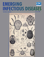
Up Close with Ticks [PDF - 1.37 MB - 2 pages]
| EID | Breedlove B. Up Close with Ticks. Emerg Infect Dis. 2023;29(1):229-230. https://doi.org/10.3201/eid2901.ac2901 |
|---|---|
| AMA | Breedlove B. Up Close with Ticks. Emerging Infectious Diseases. 2023;29(1):229-230. doi:10.3201/eid2901.ac2901. |
| APA | Breedlove, B. (2023). Up Close with Ticks. Emerging Infectious Diseases, 29(1), 229-230. https://doi.org/10.3201/eid2901.ac2901. |


