Highly Pathogenic Avian Influenza A(H5N1) Virus Clade 2.3.4.4b Infections in Wild Terrestrial Mammals, United States, 2022
Elizabeth J. Elsmo

, Arno Wünschmann, Kimberlee B. Beckmen, Liam E. Broughton-Neiswanger, Elizabeth L. Buckles, Jayne Ellis, Scott D. Fitzgerald, Robert Gerlach, Shawna Hawkins, Hon S. Ip, Julia S. Lankton, Erin M. Lemley, Julianna B. Lenoch, Mary L. Killian, Kristina Lantz, Lindsey Long, Roger Maes, Marta Mainenti, Julie Melotti, Megan E. Moriarty, Shotaro Nakagun, Rachel M. Ruden, Valerie Shearn-Bochsler, Danielle Thompson, Mia K. Torchetti, Arnaud J. Van Wettere, Annabel G. Wise, and Ailam L. Lim
Author affiliations: University of Wisconsin–Madison School of Veterinary Medicine, Madison, Wisconsin, USA (E.J. Elsmo, S. Hawkins, E.M. Lemley, A.L. Lim); Wisconsin Veterinary Diagnostic Laboratory, Madison (E.J. Elsmo, A.L. Lim); Minnesota Veterinary Diagnostic Laboratory, St. Paul, Minnesota, USA (A. Wünschmann); Alaska Department of Fish and Game, Fairbanks, Alaska, USA (K.B. Beckmen); Washington Animal Disease Diagnostic Laboratory, Pullman, Washington, USA (L.E. Broughton-Neiswanger); New York State Animal Health Diagnostic Center, Ithaca, New York, USA (E.L. Buckles, S. Nakagun); Michigan State University Veterinary Diagnostic Laboratory, Lansing, Michigan, USA (J. Ellis, S.D. Fitzgerald, R. Maes, D. Thompson, A.G. Wise); Alaska Department of Environmental Conservation, Anchorage, Alaska, USA (R. Gerlach); National Wildlife Health Center, Madison (H.S. Ip, J.S. Lankton, V. Shearn-Bochsler); Dane County Humane Society’s Wildlife Center, Madison (E.M. Lemley); US Department of Agriculture National Wildlife Research Center, Fort Collins, Colorado, USA (J.B. Lenoch); US Department of Agriculture Animal and Plant Health Inspection Service, Ames, Iowa, USA (M.L. Killian, K. Lantz, M.K. Torchetti); Wisconsin Department of Natural Resources, Madison (L. Long); Iowa State University Veterinary Diagnostic Laboratory, Ames (M.E. Mainenti, R.M. Ruden); Michigan Department of Natural Resources, Lansing (J. Melotti, M.E. Moriarty); Iowa Department of Natural Resources, Ames (R.M. Ruden); Utah Veterinary Diagnostic Laboratory, Logan, Utah, USA (A.J. Van Wettere)
Main Article
Figure 3
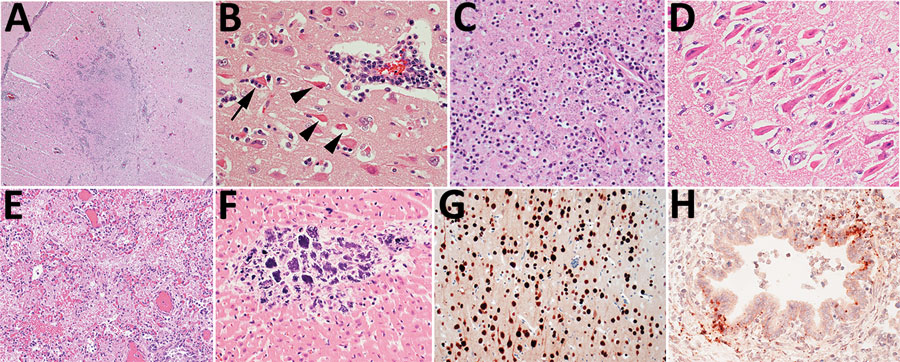
Figure 3. Histopathology of lesions in red foxes naturally infected with highly pathogenic avian influenza virus, United States. A) Throughout the brain, there are multifocal regions of necrosis and hypercellularity. Original magnification ×4. B) Within the gray matter, there is prominent neuronal necrosis (arrowheads), satellitosis (arrow), and reactive astrocytes. A vessel is surrounded by lymphocytes and plasma cells. Original magnification ×4. C) In areas of necrosis within the brain, there is often abundant, stippled, basophilic karyorrhectic debris. Original magnification ×40. D) Within the hippocampus, there are numerous shrunken, angular, and acidophilic (necrotic) neurons in a laminar pattern. Original magnification ×20. E) Within the lung, there is diffuse vascular congestion. Alveoli contain fibrin, hemorrhage, and edema fluid. Original magnification ×20. F) Regions of cardiomyocyte necrosis in the heart are often mineralized. Original magnification ×40. Panels A‒F, hematoxylin and eosin stain.G) Within the brain, there is positive nuclear and cytoplasmic staining of neuron cell bodies and processes. Avian influenza virus monoclonal immunohistochemical analysis. Original magnification ×40. H) Scattered positive nuclear and cytoplasmic staining of bronchiolar epithelial cells and interstitial macrophages in the lung. Monoclonal immunohistochemical analysis of influenza A virus nucleoprotein. Original magnification ×40.
Main Article
Page created: October 31, 2023
Page updated: November 18, 2023
Page reviewed: November 18, 2023
The conclusions, findings, and opinions expressed by authors contributing to this journal do not necessarily reflect the official position of the U.S. Department of Health and Human Services, the Public Health Service, the Centers for Disease Control and Prevention, or the authors' affiliated institutions. Use of trade names is for identification only and does not imply endorsement by any of the groups named above.