Volume 29, Number 12—December 2023
Research Letter
Human Taenia martis Neurocysticercosis, Switzerland
Abstract
Neurocysticercosis is almost exclusively caused by Taenia solium tapeworms. We describe a case of neurocysticercosis in Switzerland caused by infection with Taenia martis, the marten tapeworm, and review all 5 published cases of human infection with the marten tapeworm. In epidemiologically nonplausible cases of neurocysticercosis, zoonotic spillover infections should be suspected.
Neurocysticercosis is a zoonotic, parasitic, central nervous system infection almost exclusively caused by the larvae of Taenia solium, the pork tapeworm (1). In a few exceptional cases, neurocysticercosis in humans is not caused by T. solium but by other zoonotic Taenia species, representing rare spillover infections from distant ecologic niches (Appendix Table). In this article, we describe a rare case of T. martis neurocysticercosis in a woman in Switzerland.
A woman 55 years of age sought care at the emergency department of the Cantonal Hospital of Lucerne (Lucerne, Switzerland) because of a 3-week history of progressive transient numbness and convulsions of her left hand. Her medical history was unremarkable. Clinical examination revealed disorientation to time, a pronator drift of the left arm, hypoesthesia of the left extremities, and a tactile neglect toward the left side. A comprehensive metabolic panel and a complete blood count showed no major abnormalities. Computed tomography of the brain revealed a 12 × 14 mm mass in the right postcentral gyrus with strong ring-enhancement and perifocal edema (Appendix Figure 1). We started the patient on levetiracetam and admitted her for further investigation.
Results of computed tomography of the thorax and abdomen were unremarkable. Magnetic resonance imaging (MRI) (Appendix Figure 2) showed no restriction on diffusion-weighted imaging. After starting the patient on dexamethasone, we removed the lesion through a right parietal craniotomy. Postoperative MRI confirmed complete resection. We gradually discontinued dexamethasone and discharged the patient 6 days after the operation.
Figure
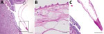
Figure. Histologic sections of resected Taenia martismetacestode from a patient in Switzerland. A) Cross-section through the excised tissue revealed strong infiltration surrounding a structure suggestive of parasitic origin. Boxed...
The specimen consisted of a cyst with a thick wall and with no macroscopically discernible content. Histologic analysis revealed a singular membrane, compatible with a helminthic parasite, and an abscessing inflammation at the border (Figure). Results of immunohistochemical tests, eubacterial 16S PCR, and whole-genome sequencing were negative, as was serologic screening for Echinococcus spp. However, Western blot for cysticercosis, which uses T. solium IgG (LDBIO Diagnostics, https://www.ldbiodiagnostics.com), revealed a weak band pattern suggestive of an infection with T. solium. We contacted the Swiss Tropical and Public Health Institute, which raised the issue of missing epidemiologic plausibility, given that the patient had never traveled to a T. solium–endemic area. The possibility of zoonotic spillover infection was considered and MRI of the whole spine, ocular ultrasound, and stool investigations recommended to exclude additional lesions and taeniasis. We analyzed the resected neurocysticercus by using a pan-helminthic PCR and sequencing of the cestode- and nematode-specific cytochrome c oxidase subunit 1 (cox1) gene (2), which revealed 100% sequence identity with Taenia martis, the marten tapeworm (GeneBank accession no. OQ536306).
Because of the unclear date of infection, the unknown proliferation rate of the parasite in humans, and the possibility of undetectable lesions, we treated the patient with albendazole and praziquantel, analogous to treatment for T. solium neurocysticercosis, assuming similar susceptibility of the parasite (3). Three months after the operation, the patient showed no neurologic deficits and no new focal seizures.
The definitive hosts of T. martis tapeworms are martens and other mustelids. T. martis tapeworms have been found in the intestines of 36% of stone martens (Martes foina) in southwest Germany but also parasitize other mustelids (4,5). Stone martens occur throughout Europe and Central Asia. The natural intermediate hosts of T. martis tapeworms are small rodents, which develop cysticerci in the pleural and peritoneal cavities. The infection in this patient suggests an accidental fecal–oral transmission, although it remains unclear when and how she came into contact with marten droppings.
Diagnosed human infections with T. martis tapeworms are limited to 5 cases reported from Germany and France (Table; Appendix Figure 3) (6–10). Those cases were 2 peritoneal infections, 2 eye infections, and 1 central nervous system infection, all with single lesions occurring in immunocompetent women. Of note, an increased susceptibility to various Taenia spp. tapeworms dependent on sex and hormone status has been described in animals (5). All 6 patients, including ours, lived in rural villages, 5 (including ours) grew their own vegetables, and at least 3 (including ours) had frequent marten sightings around their homes. Similar to what other authors reported, we did find cross-reactivity with a T. solium–specific assay.
All but 1 of the patients described in the published cases received antiparasitic treatment; the only untreated case was in a patient who refused medical treatment (10). None of the published cases reported recurrence or emergence of additional lesions.
Neurocysticercosis may be caused by Taenia spp. other than T. solium tapeworms. Cases in which an infection with T. solium is epidemiologically not plausible should be investigated for zoonotic spillover infections (in Central Europe, specifically infection with T. martis should be considered). Such cases are probably underrepresented in the literature, given the high prevalence of stone martens, their high infection rate with T. martis tapeworms, and the possibility of false positives of available, cross-reactive T. solium– and Echinococcus spp.–specific serologic assays. If adequate material is available, a panhelminthic PCR with cox1 sequencing is highly recommended.
Mr. Steinsiepe is a resident at the Department of Neurosurgery at Lucerne Cantonal Hospital, Switzerland. His primary research interest is the interface of general medicine, clinical neurology, and neurosurgery.
References
- Nash TE, Garcia HH. Diagnosis and treatment of neurocysticercosis. Nat Rev Neurol. 2011;7:584–94. DOIPubMedGoogle Scholar
- Bowles J, Blair D, McManus DP. Genetic variants within the genus Echinococcus identified by mitochondrial DNA sequencing. Mol Biochem Parasitol. 1992;54:165–73. DOIPubMedGoogle Scholar
- Neumayr A. Antiparasitic treatment recommendations: a practical guide to clinical parasitology. 2018 [cited 2022 Dec 18]. https://edoc.unibas.ch/69153
- Loos-Frank B, Zeyhle E. The intestinal helminths of the red fox and some other carnivores in southwest Germany. Z Parasitenkd. 1982;67:99–113. DOIPubMedGoogle Scholar
- Deplazes P, Eichenberger RM, Grimm F. Wildlife-transmitted Taenia and Versteria cysticercosis and coenurosis in humans and other primates. Int J Parasitol Parasites Wildl. 2019;9:342–58. DOIPubMedGoogle Scholar
- Eberwein P, Haeupler A, Kuepper F, Wagner D, Kern WV, Muntau B, et al. Human infection with marten tapeworm. Emerg Infect Dis. 2013;19:1152–4. DOIPubMedGoogle Scholar
- Brunet J, Benoilid A, Kremer S, Dalvit C, Lefebvre N, Hansmann Y, et al. First case of human cerebral Taenia martis cysticercosis. J Clin Microbiol. 2015;53:2756–9. DOIPubMedGoogle Scholar
- Koch T, Schoen C, Muntau B, Addo M, Ostertag H, Wiechens B, et al. Molecular diagnosis of human Taenia martis eye infection. Am J Trop Med Hyg. 2016;94:1055–7. DOIPubMedGoogle Scholar
- Rudelius M, Brehm K, Poelcher M, Spinner C, Rosenwald A, da Costa CP. First case of human peritoneal cysticercosis mimicking peritoneal carcinosis: necessity of laparoscopy and histologic assessment for the correct diagnosis. JMM Case Rep. 2017;4:
e005097 . DOIPubMedGoogle Scholar - Mueller A, Förch G, Zustin J, Muntau B, Schuldt G, Tappe D. Case report: molecular identification of larval Taenia martis infection in the pouch of Douglas. Am J Trop Med Hyg. 2020;103:2315–7. DOIPubMedGoogle Scholar
Figure
Table
Cite This ArticleOriginal Publication Date: November 14, 2023
Table of Contents – Volume 29, Number 12—December 2023
| EID Search Options |
|---|
|
|
|
|
|
|
Please use the form below to submit correspondence to the authors or contact them at the following address:
Valentin K. Steinsiepe, Department of Neurosurgery, Cantonal Hospital of Lucerne, Spitalstrasse, 6000 Luzern, Switzerland
Top