Skip directly to site content Skip directly to page options Skip directly to A-Z link Skip directly to A-Z link Skip directly to A-Z link
Volume 29, Number 6—June 2023
Dispatch

MERS-CoV‒Specific T-Cell Responses in Camels after Single MVA-MERS-S Vaccination

Christian Meyer zu Natrup, Lisa-Marie Schünemann, Giulietta Saletti, Sabrina Clever, Vanessa Herder, Sunitha Joseph, Marina Rodriguez, Ulrich Wernery, Gerd Sutter, and Asisa VolzComments to Author 
Author affiliations: University of Veterinary Medicine Hannover, Hanover, Germany (C. Meyer zu Natrup, L.-M. Schünemann, G. Saletti, S. Clever, A. Volz); University of Glasgow, Glasgow, Scotland, UK (V. Herder); Central Veterinary Research Laboratory, Dubai, United Arab Emirates (S. Joseph, M. Rodriguez, U. Wernery); Ludwig Maximilian University Munich, Munich, Germany (G. Sutter)

Main Article

Figure 1

Antigen-specific humoral immunity after MVA-MERS-S vaccination in dromedary camels, Dubai, United Arab Emirates. MERS-CoV seropositive and naive dromedary camels were immunized once with 2.5 x 10⁸ plaque-forming units of MVA-MERS-S or MVA as a vector control. Serum samples were collected on day 0 and on day 15 after single-shot vaccination. Black indicates serum samples analyzed for MERS-CoV S1 IgG by ELISA of MVA–vaccinated animals and red indicates for MVA-MERS-S–vaccinated animals. Box plots show individual values (dots), median values (horizontal lines within boxes), first and third quartiles (box tops and bottoms), and minimums and maximums of value distribution (error bars). LOD, limit of detection; MERS-CoV, Middle East respiratory syndrome coronavirus; MVA, modified vaccinia virus Ankara; MVA-MERS-S, modified vaccinia virus Ankara expressing full-length MERS-CoV spike protein as antigen.

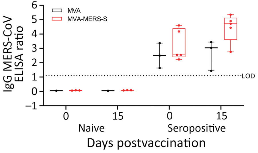
Figure 1. Antigen-specific humoral immunity after MVA-MERS-S vaccination in dromedary camels, Dubai, United Arab Emirates. MERS-CoV seropositive and naive dromedary camels were immunized once with 2.5 x 10⁸ plaque-forming units of MVA-MERS-S or MVA as a vector control. Serum samples were collected on day 0 and on day 15 after single-shot vaccination. Black indicates serum samples analyzed for MERS-CoV S1 IgG by ELISA of MVA–vaccinated animals and red indicates for MVA-MERS-S–vaccinated animals. Box plots show individual values (dots), median values (horizontal lines within boxes), first and third quartiles (box tops and bottoms), and minimums and maximums of value distribution (error bars). LOD, limit of detection; MERS-CoV, Middle East respiratory syndrome coronavirus; MVA, modified vaccinia virus Ankara; MVA-MERS-S, modified vaccinia virus Ankara expressing full-length MERS-CoV spike protein as antigen.

Main Article

Page created: March 29, 2023
Page updated: May 17, 2023
Page reviewed: May 17, 2023
The conclusions, findings, and opinions expressed by authors contributing to this journal do not necessarily reflect the official position of the U.S. Department of Health and Human Services, the Public Health Service, the Centers for Disease Control and Prevention, or the authors' affiliated institutions. Use of trade names is for identification only and does not imply endorsement by any of the groups named above.
file_external