Microscopic Evidence of Malaria Infection in Visceral Tissue from Medici Family, Italy
Frank Maixner, Dennis Drescher, Giulia Boccalini, Dario Piombino-Mascali, Marek Janko, Nicole Berens-Riha, Bum Jin Kim, Michelle Gamble, Jolanthe Schatterny, Rory E. Morty, Melanie Ludwig, Ben Krause-Kyora, Robert Stark, Hyun Joo An, Jens Neumann, Giovanna Cipollini, Rudolf Grimm, Nicole Kilian, and Albert Zink

Author affiliations: Eurac Research, Bolzano, Italy (F. Maixner, G. Boccalini, G. Cipollini, A. Zink); University Hospital Heidelberg, Heidelberg, Germany (D. Drescher, J. Schatterny, R.E. Morty, M. Ludwig, N. Kilian); Vilnius University, Vilnius, Lithuania (D. Piombino-Mascali); Technical University of Darmstadt, Darmstadt, Germany (M. Janko, R. Stark); Institute of Tropical Medicine, Antwerp, Belgium (N. Berens-Riha); University of Munich, Munich, Germany (N. Berens-Riha); Oregon State University, Corvallis, Oregon, USA (B.J. Kim); Chungnam National University, Daejeon, Korea (B.J. Kim, H.J. An); Heritage and Archaeological Research Practice, Edinburgh, Scotland, UK (M. Gamble); Kiel University, Kiel, Germany (B. Krause-Kyora); Ludwig Maximilian University, Munich (J. Neumann); Agilent Technologies, Santa Clara, California, USA (R. Grimm)
Main Article
Figure
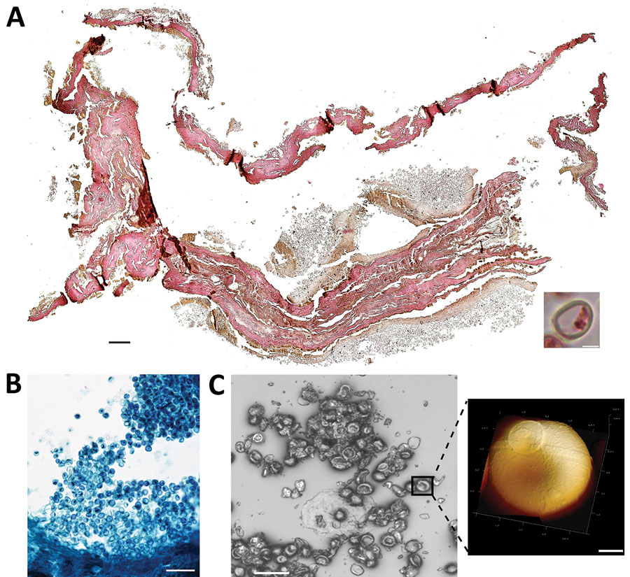
Figure. Microscopic analysis of malaria infection in visceral tissue from Medici family, Italy. We evaluated a 2.5 cm × 1.5 cm tissue piece (ID 1297) from 1 jar containing viscera of a Medici family member and identified a potential blood vessel containing erythrocytes. A) Histological cross section of the tissue stained with hematoxylin and eosin; scale bar indicates 200 µm. Inset shows a possible erythrocyte; scale bar indicates 3 µm. B) Giemsa staining of a paraffin section of viscera suggesting the presence of parasites within the erythrocytes. Scale bar indicates 50 µm. C) Atomic force microscopy (AFM) of the tissue section. An optical microscope was used to define appropriate sample areas for AFM imaging (left image); scale bar indicates 20 µm. Enlarged area at right shows a ring stage of Plasmodium falciparum in an erythrocyte; scale bar indicates 2 µm.
Main Article
Page created: April 12, 2023
Page updated: May 18, 2023
Page reviewed: May 18, 2023
The conclusions, findings, and opinions expressed by authors contributing to this journal do not necessarily reflect the official position of the U.S. Department of Health and Human Services, the Public Health Service, the Centers for Disease Control and Prevention, or the authors' affiliated institutions. Use of trade names is for identification only and does not imply endorsement by any of the groups named above.