Volume 29, Number 8—August 2023
Research
Elimination of Dirofilaria immitis Infection in Dogs, Linosa Island, Italy, 2020–2022
Abstract
On Linosa Island, Italy, Dirofilaria immitis infection has been hyperendemic in dogs and seroprevalent among islanders. In 2020, we implemented a heartworm disease elimination program on Linosa Island. Of 54 dogs tested for D. immitis antigen and microfilariae, 28 had positive results and received treatment with oral doxycycline twice daily for 4 weeks plus topical imidacloprid/moxidectin monthly for 12 months. The 26 dogs with negative results received monthly topical imidacloprid/moxidectin as preventive. During month 1, the number of microfilaremic dogs was reduced by 76.5%. From month 2 on, all animals were microfilariae negative, and during months 3 to 9, the number of antigen-positive dogs decreased progressively. Treatment of positive dogs coupled with chemoprophylaxis for noninfected dogs was effective, protecting them from new infections. The elimination program reduced the risk for human infection, representing a One Health paradigm. Monitoring and chemoprophylaxis are advocated to maintain the status of heartworm disease–free area.
Dirofilaria immitis and D. repens (Spirurida, Onchocercidae) nematodes are among the most common species of filariae that cause diseases in dogs and other animals; both species infect humans (1,2). In dogs, D. immitis filariae cause the severe illness heartworm disease (HWD), whereas D. repens infection is less severe. Both Dirofilaria species are transmitted by mosquitoes; in Europe, the competent vectors are Culex pipiens and Aedes albopictus mosquitoes (3). However, the involvement of flying insects other than mosquitos has been recently hypothesized, including black flies belonging to the Simulium turgaicum complex (4) and Culicoides paolae biting midges (5).
Unlike D. repens filariae, which are more widely distributed in Europe, including the Italian Peninsula (1,6–8), D. immitis worms are more frequently recorded in central Europe (9), including regions in northern Italy (10,11). Nevertheless, autochthonous cases of HWD in dogs from central and southern Italy have been retrospectively reported from 2009 (1) through 2019 (12); highly endemic foci in southern regions of Italy have been described (13,14). That new epidemiologic scenario developed after the arrival of a new invasive mosquito species (i.e., Ae. albopictus) and the increased movement of animals throughout the country combined with lack of chemoprophylactic strategies for dogs from non–HWD-endemic regions (9,12,13). The island of Linosa, Italy, represents a paradigm of that scenario. A highly endemic focus of D. immitis filariae was recently described on the small and remote island located in the middle of the Mediterranean Basin, where 58.9% of dogs tested positive for HWD by modified Knott and SNAP 4Dx Plus tests (14). In the same epidemiologic context, 7.9% of human islanders tested positive for D. immitis antibodies (15), which emphasizes the role of D. immitis–infected dogs as a source for human infections in specifically isolated environments, thus advocating for treating infected animals in the context of One Health.
Traditionally, HWD is treated with melarsomine dihydrochloride, the sole registered heartworm adulticide drug (16,17). However, an alternative therapeutic approach, known as the slow-kill protocol, that combines macrocyclic lactones with doxycycline, targeting the bacterial endosymbiont Wolbachia, has been used successfully in experimentally and naturally infected dogs, (18–20). That protocol was recently recognized by the American Heartworm Society and the European Society of Dirofilariosis and Angiostrongylosis as an alternative strategy when treatment with melarsomine is either unavailable or contraindicated. Compared with the melarsomine treatment, doxycycline (10 mg/kg 2×/d) for 4 weeks combined with monthly administration of a topical formulation of 10% wt/vol imidacloprid and 2.5% wt/vol moxidectin for 9 months proved to be safe and effective for treating HWD in naturally infected dogs and for clearing dogs of circulating microfilariae (20).
Because of its geographic and epidemiologic peculiarities, Linosa Island offered an exceptional opportunity to use this elimination program for HWD. The program involved therapeutically and prophylactically administering the alternative protocol to all infected dogs and administering monthly preventive of 10% wt/vol imidacloprid plus 2.5% wt/vol moxidectin to all remaining uninfected dogs on the island. The study was complemented by an entomologic survey to assess mosquito vectors within this unique epidemiologic context.
The study was approved by the ethics committee of animal experiments of the Department of Veterinary Medicine, University of Bari, Italy (approval number 01/2021). It was conducted according to the VICH GL9 principles of Good Clinical Practice (21).
Animal Sampling and Diagnosis
In October 2020 (T0), we physically examined all 58 dogs on Linosa Island and recorded signalment and history (e.g., age, sex, breed, clinical signs, and treatments) in individual files. At T0, we collected blood samples from each dog and stored them in two 1-mL K3EDTA tubes and in one 5-mL tube with clot activator. We used the Knott test (22) to detect and identify circulating microfilariae in whole-blood samples and a duplex real-time quantitative PCR (qPCR) to differentiate Dirofilaria species (23).
We analyzed serum samples for the presence of D. immitis–specific antigens by using the SNAP 4Dx Plus rapid ELISA (IDEXX, https://www.idexx.com). We considered dogs that were positive for D. immitis, either microscopically, serologically, or molecularly, to be infected and assigned them to the treatment group (G1); we assigned dogs that were negative to the prevention group (G2). Before the beginning of treatment, we performed cardiac ultrasonography for all D. immitis–infected dogs to detect adult parasites in the pulmonary arteries. We used an echocardiographic unit (Vivid-iQ; GE Healthcare, https://www.gehealthcare.com) equipped with dedicated multifrequency phased array transducers (6s-RS and M5-RS). In brief, we used the right parasternal long-axis view optimized for the right pulmonary vein and artery (standard view 1) and the right parasternal short-axis view optimized for the right pulmonary artery (standard view 2) to detect worms (24). We did not perform thoracic radiography because no facility was available on the island.
Treatment and Follow-up
D. immitis–infected dogs (G1) received doxycycline (Ronaxan; Boehringer Ingelheim, https://www.boehringer-ingelheim.com) (10 mg/kg orally 2×/d) for 4 weeks plus a monthly application of a spot-on formulation containing 10% wt/vol imidacloprid and 2.5% wt/vol moxidectin (Advocate; Elanco Animal Health, www.elanco.com) for 12 months. Dogs in the G2 group underwent monthly chemoprophylactic treatment with the same spot-on product used for the G1 dogs.
Figure 1
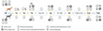
Figure 1. Schematic design of study of elimination of Dirofilaria immitisinfection in dogs, Linosa Island, Italy, 2020, and follow-up examinations. G1, infected group; G2, noninjected group; T, time after start...
G1 dogs underwent follow-up examination at 1, 2, 3, 6, 9, 12 and 18 months (designated as T1, T2, T3, T6, T9, T12, and T18) after enrollment (Figure 1). At each follow-up visit, we clinically examined dogs and collected blood samples that we analyzed by Knott test (at T1, T2, and T18) or by SNAP 4Dx Plus test (at T1, T2, T3, T6, T9, T12 and T18). We repeated cardiac ultrasonography for G1 dogs at T3, T6, T9, T12, and T18. G2 dogs underwent clinical examination monthly, and we tested them for parasite antigenemia (by SNAP 4Dx Plus test) at T3, T6, T9, T12, and T18 (Figure 1).
Of the 58 dogs examined at T0, we included only 54 in the study because 2 dogs were going to be on the island for only a few weeks/months and the owners of the other 2 dogs did not consent to their study participation. For dogs that were permanently introduced onto the island after the beginning of the study, we performed clinical examination and specific testing (i.e., Knott test and testing for D. immitis circulating antigens) with owner consent and allocated them to G1 or G2 according to the test results. We advised owners to immediately report any clinical signs (i.e., cough, hemoptysis, syncope) that might appear during the study and to reduce physical activity for all infected dogs, regardless of symptoms, as much as possible.
Mosquito Collection
Figure 2
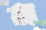
Figure 2. Linosa Island (Sicily, Italy) indicating the positions of infected dogs, noninfected dogs, and light traps used to capture mosquitoes. Inset shows location of Linosa Island.
We collected mosquitos from 5 locations where cases of D. immitis infection in dogs were detected (Figure 2). We trapped mosquitoes daily during October 2020–November 2021, 5:30 p.m.–9:00 a.m., by using 1 light trap per site, set ≈50 cm above the ground. We replaced net bags daily and stored insects in the net bags at −20°C according to collection site/day. After morphologically identifying mosquitoes to the species level (25), we further analyzed female mosquitoes with qPCR to detect and differentiate Dirofilaria spp.
Molecular Diagnosis
We extracted genomic DNA from samples of dog blood by using the commercial QIAamp DNA Micro Kit (QIAGEN GmbH, https://www.qiagen.com) and samples of mosquito abdomen and thorax by using an in-house method (26). We tested all DNA samples with qPCR by using 2 species-specific primer sets targeting partial cytochrome c oxidase subunit 1 for D. immitis DNA and the second internal transcribed spacer-2 of nuclear ribosomal DNA for D. repens DNA, as previously described (23). We tested all DNA samples in duplicate and included positive and negative controls in each qPCR run.
We detected D. immitis infection in 28 (51.9%) of 54 (33 male, 21 female) enrolled dogs (Appendix). We found circulating microfilariae in 17 (60.7%) infected dogs and specific D. immitis antigens in 27 (96.4%). All microfilaremic animals except 1 had positive ELISA antigen test results (Appendix). Microfilariae were morphologically and molecularly identified as D. immitis.
At the time of enrollment (T0), we conducted echocardiography on 23 of 28 infected dogs and found D. immitis adults within the pulmonary arteries of 20 (Appendix). On T0, we allocated 28 dogs to G1 and 26 to G2. A total of 4 dogs (3 in G1 and 1 in G2) were unavailable for follow-up analysis because of events not associated with treatment (e.g., low owner compliance or neoplastic disease). During the study period, 5 new dogs were permanently introduced onto the island (3 in January 2021 and 2 in March 2021); they tested negative for D. immitis infection and were included in group G2.
At T1, the number of microfilaremic dogs was reduced by 76.5%; only 4 of 17 dogs were positive by Knott test. From T2 until the end of the study, no microfilariae were found in G1 dogs. By T1, 3 infected dogs had become antigen negative (no antigen detected) according to ELISA test (Appendix). From T3 on, the number of antigen-positive dogs decreased progressively (20 at T3, 10 at T6, and 2 at T9) (Appendix). All dogs in G1 were negative for D. immitis circulating antigens at the 1-year follow-up visit (T12, October 2021).
Cardiac ultrasonography showed filarial parasites in the pulmonary arteries in 14 dogs at T3, 9 dogs at T6, 6 dogs at T9, and 3 dogs at T12 (Appendix). At T18, all G1 dogs scored negative by Knott test and ELISA antigen test, and no parasites were detected by echocardiography. Group 2 dogs tested negative for D. immitis antigens at all scheduled follow-up visits.
In this study, treatment with doxycycline and 10% wt/vol imidacloprid plus 2.5% wt/vol moxidectin seemed to be safe, and no adverse events were observed. However, in 1 dog from the G1 group, a thromboembolic-like disorder with hindlimb paralysis and pain was observed a few days after the beginning of treatment. Doxycycline treatment was temporarily suspended, and fluid therapy was promptly administered in association with unfractionated heparin (200 U/kg subcutaneously every 8 h for 3 d) and prednisone (1 mg/kg 2×/d in gradually decreasing doses for 15 d), resulting in full recovery of that G1 animal after 5 days.
Another dog that was apparently healthy at T0 exhibited ascites and diffuse edema of the hindlimbs at the clinical examination conducted on T3. Ultrasonography revealed a high parasite burden within the pulmonary arteries, associated with posthepatic portal hypertension. Treatment with sildenafil (2 mg/kg 3×/d for >6 mo) was administered. At the T6 follow-up visit, the dog had improved clinically, although pulmonary hypertension was still present, but pulmonary hypertension was absent at the next follow-up visits.
During this study, we collected 359 mosquitoes (169 male and 190 female) belonging to 6 species, the most abundant of which was C. pipiens (Table). Of the 190 females collected, 53 were engorged, and none of the analyzed specimens scored positive to Dirofilaria spp. DNA.
In a short time, D. immitis parasites were eliminated from Linosa Island after we combined an alternative adulticide protocol for infected dogs with administration of preventives to noninfected animals. The peculiar epidemiologic context of the island (i.e., geographically confined area, limited number of dogs, and absence of wild reservoirs) aided the campaign. After the HWD focus in southernmost Europe was described (14), we planned and successfully implemented our project. In contrast to programs targeting other mosquitoborne diseases, such as malaria, this elimination campaign did not target the vector (27) and instead targeted the parasite specifically by eliminating the circulating juvenile forms and adult parasites in infected dogs while simultaneously protecting noninfected animals with chemoprophylaxis.
Treatment with doxycycline and imidacloprid/moxidectin was effective and safe for all of the dogs; side effects were observed in only 2 dogs, probably resulting from thromboembolism after worm death, which is a side effect associated with any adulticide protocol. Nevertheless, the risk for thromboembolism is also linked to patients’ excessive physical activity after treatment, which cannot be ruled out (28). Furthermore, protocols that cause the progressive, slow death of adult parasites expand the temporal window of thromboembolic risk compared with conventional treatment (24). A retrospective study suggests that the risk for thromboembolic events is higher 3 months after the start of the alternative treatment protocol than after start of the melarsomine protocol and that exercise restriction for treated animals is advisable for longer periods or until antigen test results are negative (28,29). Regardless, the alternative protocol has proven to be effective (20,24,30) and may result in fewer adverse events than those resulting from melarsomine alone (31,32) if exercise restriction and other precautions are adequately implemented.
For this study, we chose the combined doxycycline/moxidectin protocol over the conventional adulticide therapy, considering the potential adverse events associated with the sudden death of adult worms causing pulmonary thrombosis (32) and the lack of veterinary services available on the island. On the other hand, the adulticide effects of doxycycline on developing and adult stages of the parasite are slow (33), and macrocyclic lactones are effective against D. immitis L3 and L4 and kill only adult parasites after prolonged use (2). Whether the 2 drugs work better together to eliminate D. immitis parasites in a short time because of a cumulative or synergistic effect is not known.
Eliminating circulating microfilariae in infected dogs after the beginning of the study (T2) drastically reduced the number of dogs acting as reservoirs on the island and therefore the risk for D. immitis infection of mosquitoes. All of the actions contributed to breaking the life cycle of the parasite, a finding that was supported by the fact that none of the examined mosquitoes scored positive for Dirofilaria spp. DNA up to November 2021, unlike before the study had started a few months earlier (14).
The protocol that we used led to progressive reduction of detectable antigens in treated animals from T3 to T9 and to dogs testing negative at T12. This finding is similar or even better than what was recorded in previous studies that used the same protocol (20,34,35). Also, the conversion to the antigen-negative status observed was faster than that recorded with ivermectin alone (24,36) or with ivermectin (every 2 weeks for 6 months) and doxycycline (10 mg/kg/d orally for 30 d) in which only 73% or 80% of treated dogs reached antigen-negative status (30,31).
The use of macrocyclic lactones as adulticides has been discouraged by several authors because it may take many months for adult worms to die, enabling the disease to progress and allowing for the potential selection of macrocyclic lactone–resistant strains (37). Moreover, potential interference with antigen test results has also been suggested (37). Several studies have reported false-negative antigen test results in dogs and cats treated with macrocyclic lactones, presumably resulting from formation of immune complexes and the consequent binding of the antigens (37–39). To overcome this problem, 2 consecutive negative antigen test results 6 months apart have been suggested as a valid criterion for considering a dog cleared of infection (34). Therefore, in this elimination program, all dogs were probably cured of infection because in no dog was antigen detected at T12 or 6 months after (T18). This finding is also strongly supported by ultrasonography in which no adult heartworms were observed at the T18 follow-up visit. Our data may also be useful in where D. immitis parasites are endemic and melarsomine is not commercially available (e.g., Brazil, eastern Europe).
Despite the presence of domestic and feral cats on Linosa Island (40), our study did not include those hosts because of lack of owner compliance and the difficulty of sampling these animals in a remote environment. However, considering the occurrence and possible role of cats as reservoirs of D. immitis parasites (41), further studies evaluating the prevalence of this parasite in cat populations on Linosa Island are warranted.
Considering the zoonotic potential of D. immitis worms, elimination of HWD in dogs is pivotal for reducing the risk for human infection. Indeed, in previous surveys conducted on the human population from Linosa Island, up to 7.9% of islanders were positive for circulating D. immitis antibodies (15), which represented one of the highest percentages of human exposure ever reported. Therefore, results from our study suggest that adulticide treatment of all infected dogs, coupled with prevention for noninfected dogs, not only protects animals from HWD but may also potentially reduce the risk for human infection. Such an approach represents a paradigm for the One Health concept. Continuous monitoring and chemoprophylaxis of the canine population on the island and of all newly introduced dogs are highly recommended to maintain the status of HWD-free area.
Dr. Brianti is full professor at Department of Veterinary Sciences, University of Messina, Italy. His main research activity is focused on parasitic diseases of veterinary and public health concern.
Acknowledgments
We thank Laura Helen Kramer for her critical revision of the manuscript and acknowledge Elanco Animal Health for financial support of this study.
The study was conducted under the frame of the Project PE-13, INF-ACT, which is part of the National Recovery and Resilience Plan.
References
- Otranto D, Capelli G, Genchi C. Changing distribution patterns of canine vector borne diseases in Italy: leishmaniosis vs. dirofilariosis. Parasit Vectors. 2009;2(Suppl 1):S2. DOIPubMedGoogle Scholar
- McCall JW, Genchi C, Kramer LH, Guerrero J, Venco L. Heartworm disease in animals and humans. Adv Parasitol. 2008;66:193–285. DOIPubMedGoogle Scholar
- Eldridge BF, Edman JDI. Introduction to medical entomology. In: Eldridge BF, Edman JD, editors. Medical Entomology. Berlin: Springer; 2000. p. 1–13.
- Khanzadeh F, Khaghaninia S, Maleki-Ravasan N, Koosha M, Oshaghi MA. Molecular detection of Dirofilaria spp. and host blood-meal identification in the Simulium turgaicum complex (Diptera: Simuliidae) in the Aras River Basin, northwestern Iran. Parasit Vectors. 2020;13:548. DOIPubMedGoogle Scholar
- Napoli E, Panarese R, La Russa F, Cambera I, Mendoza-Roldan JA, Otranto D, et al. Detection of Dirofilaria DNA and host blood-meal identification in Culicoides paolae biting-midges. Parasitology. 2022;149:1–17. DOIPubMedGoogle Scholar
- Capelli G, Genchi C, Baneth G, Bourdeau P, Brianti E, Cardoso L, et al. Recent advances on Dirofilaria repens in dogs and humans in Europe. Parasit Vectors. 2018;11:663. DOIPubMedGoogle Scholar
- Fioretti DP, Diaferia M, Grelloni V, Maresca C. Canine filariosis in Umbria: an update of the occurrence one year after the first observation of autochthonous foci. Parassitologia. 2003;45:79–83.PubMedGoogle Scholar
- Scaramozzino P, Gabrielli S, Di Paolo M, Sala M, Scholl F, Cancrini G. Dog filariosis in the Lazio region (Central Italy): first report on the presence of Dirofilaria repens. BMC Infect Dis. 2005;5:75. DOIPubMedGoogle Scholar
- Genchi C, Kramer LH. The prevalence of Dirofilaria immitis and D. repens in the Old World. Vet Parasitol. 2020;280:
108995 . DOIPubMedGoogle Scholar - Genchi C, Rinaldi L, Cascone C, Mortarino M, Cringoli G. Is heartworm disease really spreading in Europe? Vet Parasitol. 2005;133:137–48. DOIPubMedGoogle Scholar
- Genchi C, Rinaldi L, Mortarino M, Genchi M, Cringoli G. Climate and Dirofilaria infection in Europe. Vet Parasitol. 2009;163:286–92. DOIPubMedGoogle Scholar
- Mendoza-Roldan J, Benelli G, Panarese R, Iatta R, Furlanello T, Beugnet F, et al. Leishmania infantum and Dirofilaria immitis infections in Italy, 2009-2019: changing distribution patterns. Parasit Vectors. 2020;13:193. DOIPubMedGoogle Scholar
- Panarese R, Iatta R, Mendoza-Roldan JA, Szlosek D, Braff J, Liu J, et al. Comparison of diagnostic tools for the detection of Dirofilaria immitis infection in dogs. Pathogens. 2020;9:499. DOIPubMedGoogle Scholar
- Brianti E, Panarese R, Napoli E, De Benedetto G, Gaglio G, Bezerra-Santos MA, et al. Dirofilaria immitis infection in the Pelagie archipelago: The southernmost hyperendemic focus in Europe. Transbound Emerg Dis. 2022;69:1274–80. DOIPubMedGoogle Scholar
- Mendoza-Roldan JA, Gabrielli S, Cascio A, Manoj RRS, Bezerra-Santos MA, Benelli G, et al. Zoonotic Dirofilaria immitis and Dirofilaria repens infection in humans and an integrative approach to the diagnosis. Acta Trop. 2021;223:
106083 . DOIPubMedGoogle Scholar - Keister DM, Dzimianski MT, McTier TL, McCall JW, Brown J. Dose selection and confirmation of RM 340, a new filaricide for the treatment of dogs with immature and mature Dirofilaria immitis. In: Sol MD, editor. Proceedings of the Heartworm Symposium ’92; Austin, Texas, USA; March 7–29, 1992. p. 225–9.
- Vezzoni A, Genchi C, Raynaud JP. Adulticide efficacy of RM340 in dogs with mild and severe natural infections. In: Proceedings of the Heartworm Symposium ’92; Austin, Texas, USA; March 7–29, 1992. p. 231–240.
- Bazzocchi C, Mortarino M, Grandi G, Kramer LH, Genchi C, Bandi C, et al. Combined ivermectin and doxycycline treatment has microfilaricidal and adulticidal activity against Dirofilaria immitis in experimentally infected dogs. Int J Parasitol. 2008;38:1401–10. DOIPubMedGoogle Scholar
- Manoj RRS, Latrofa MS, Mendoza-Roldan JA, Otranto D. Molecular detection of Wolbachia endosymbiont in reptiles and their ectoparasites. Parasitol Res. 2021;120:3255–61. DOIPubMedGoogle Scholar
- Genchi M, Vismarra A, Lucchetti C, Viglietti A, Crosara S, Gnudi G, et al. Efficacy of imidacloprid 10%/moxidectin 2.5% spot on (Advocate®, Advantage Multi®) and doxycycline for the treatment of natural Dirofilaria immitis infections in dogs. Vet Parasitol. 2019;273:11–6. DOIPubMedGoogle Scholar
- European Medicines Agency/Committee for Veterinary Medicinal Products. VICH GL9 principles of good clinical practice [cited 2023 Jun 9]. https://www.ema.europa.eu/en/documents/scientific-guideline/vich-gl9-good-clinical-practices-step-7_en.pdf
- Genchi M, Ciuca L, Vismarra A, Ciccone E, Cringoli G, Kramer L, et al. Evaluation of alternative reagents on the performance of the modified Knott’s test. Vet Parasitol. 2021;298:
109555 . DOIPubMedGoogle Scholar - Latrofa MS, Annoscia G, Dantas-Torres F, Traversa D, Otranto D. Towards a rapid molecular identification of the common phlebotomine sand flies in the Mediterranean region. Vet Parasitol. 2012;184:267–70. DOIPubMedGoogle Scholar
- Venco L, Mihaylova L, Boon JA. Right Pulmonary Artery Distensibility Index (RPAD Index). A field study of an echocardiographic method to detect early development of pulmonary hypertension and its severity even in the absence of regurgitant jets for Doppler evaluation in heartworm-infected dogs. Vet Parasitol. 2014;206:60–6. DOIPubMedGoogle Scholar
- Severini F, Toma L, Di Luca M, Romi R. Le zanzare italiane: generalità e identificazione e gli adulti (Diptera, Culicidae). Fragm Entomol. 2009;41:213–372. DOIGoogle Scholar
- Latrofa MS, Angelou A, Giannelli A, Annoscia G, Ravagnan S, Dantas-Torres F, et al. Ticks and associated pathogens in dogs from Greece. Parasit Vectors. 2017;10:301. DOIPubMedGoogle Scholar
- Kumar A. Some considerable issues concerning malaria elimination in India. J Vector Borne Dis. 2019;56:25–31. DOIPubMedGoogle Scholar
- Ames MK, VanVranken P, Evans C, Atkins CE. Non-Arsenical heartworm adulticidal therapy using topical moxidectin-imidacloprid and doxycycline: A prospective case series. Vet Parasitol. 2020;282:
109099 . DOIPubMedGoogle Scholar - Ames MK, Atkins CE. Treatment of dogs with severe heartworm disease. Vet Parasitol. 2020;283:
109131 . DOIPubMedGoogle Scholar - Grandi G, Quintavalla C, Mavropoulou A, Genchi M, Gnudi G, Bertoni G, et al. A combination of doxycycline and ivermectin is adulticidal in dogs with naturally acquired heartworm disease (Dirofilaria immitis). Vet Parasitol. 2010;169:347–51. DOIPubMedGoogle Scholar
- Mavropoulou A, Gnudi G, Grandi G, Volta A, Kramer LH, Quintavalla C. Clinical assessment of post-adulticide complications in Dirofilaria immitis-naturally infected dogs treated with doxycycline and ivermectin. Vet Parasitol. 2014;205:211–5. DOIPubMedGoogle Scholar
- Kramer L, Grandi G, Passeri B, Gianelli P, Genchi M, Dzimianski MT, et al. Evaluation of lung pathology in Dirofilaria immitis-experimentally infected dogs treated with doxycycline or a combination of doxycycline and ivermectin before administration of melarsomine dihydrochloride. Vet Parasitol. 2011;176:357–60. DOIPubMedGoogle Scholar
- McCall JW. The safety-net story about macrocyclic lactone heartworm preventives: a review, an update, and recommendations. Vet Parasitol. 2005;133:197–206. DOIPubMedGoogle Scholar
- Bendas AJR, Mendes-de-Almeida F, Von Simson C, Labarthe N. Heat pretreatment of canine samples to evaluate efficacy of imidacloprid + moxidectin and doxycycline in heartworm treatment. Parasit Vectors. 2017;10:246. DOIPubMedGoogle Scholar
- Savadelis MD, Ohmes CM, Hostetler JA, Settje TL, Zolynas R, Dzimianski MT, et al. Assessment of parasitological findings in heartworm-infected beagles treated with Advantage Multi® for dogs (10% imidacloprid + 2.5% moxidectin) and doxycycline. Parasit Vectors. 2017;10:245. DOIPubMedGoogle Scholar
- McCall JW, Guerrero J, Roberts RE, Supakorndej N, Mansour AE, Dzimianski MT, et al. Further evidence of clinical prophylactic, retroactive (reach back), and adulticidal activity of monthly administration of ivermectin (Heartgard PlusTM) in dogs experimentally infected with heartworms. In: Proceedings of the Heartworm Symposium ’01; San Antonio, Texas, USA; April 20–22, 2001. p. 189–200.
- Drake J, Gruntmeir J, Merritt H, Allen L, Little SE. False negative antigen tests in dogs infected with heartworm and placed on macrocyclic lactone preventives. Parasit Vectors. 2015;8:68–73. DOIPubMedGoogle Scholar
- Velasquez L, Blagburn BL, Duncan-Decoq R, Johnson EM, Allen KE, Meinkoth J, et al. Increased prevalence of Dirofilaria immitis antigen in canine samples after heat treatment. Vet Parasitol. 2014;206:67–70. DOIPubMedGoogle Scholar
- Little SE, Munzing C, Heise SR, Allen KE, Starkey LA, Johnson EM, et al. Pre-treatment with heat facilitates detection of antigen of Dirofilaria immitis in canine samples. Vet Parasitol. 2014;203:250–2. DOIPubMedGoogle Scholar
- Ozella L, Cecchetti M, Pessani D. Diet of feral cats during the Scopoli’s shearwater breeding season on Linosa Island, Mediterranean Sea. Ital J Zool (Modena). 2016;83:589–99. DOIGoogle Scholar
- Panarese R, Iatta R, Lia RP, Passantino G, Ciccarelli S, Gernone F, et al. Dirofilarioses in two cats in southern Italy. Parasitol Res. 2021;120:4247–51. DOIPubMedGoogle Scholar
Figures
Table
Cite This ArticleOriginal Publication Date: June 21, 2023
Table of Contents – Volume 29, Number 8—August 2023
| EID Search Options |
|---|
|
|
|
|
|
|
Please use the form below to submit correspondence to the authors or contact them at the following address:
Emanuele Brianti, University of Messina, Viale Giovanni Palatucci, 98168 Messina, Italy;ebrianti@unime.it or Domenico Otranto, University of Bari Aldo Moro, strada provinciale per Casamassima km3, 70010 Valenzano, Italy; domenico.otranto@uniba.it
Top