Skip directly to site content Skip directly to page options Skip directly to A-Z link Skip directly to A-Z link Skip directly to A-Z link
Volume 30, Number 1—January 2024
Dispatch

Helicobacter fennelliae Localization to Diffuse Areas of Human Intestine, Japan

Takashi Sakoh, Emiko Miyajima, Yusuke Endo, Kei Kono, Junichiro Sato, Mizuki Haraguchi, Sho Ogura, Masayo Morishima, Keiko Ishida, Yorinari Ochiai, Shu Hoteya, Yutaka Takazawa, Masaru Baba, and Hideki AraokaComments to Author 
Author affiliation: Toranomon Hospital, Tokyo, Japan

Main Article

Figure 2

Microscopic findings in ileocolonic biopsy specimens (second colonoscopy) in a man in Japan who had recurrent Helicobacter fennelliae bacteremia. A) Morphologic features of the bacteria in a cecal tissue suspension with Gram staining (original magnification ×2,000). Arrow indicates gram-negative spiral bacilli. B) Histologic findings in a biopsy specimen taken from the transverse colon with hematoxylin-eosin staining (original magnification ×200). The colonic mucosa shows mild leukocytic infiltration. C) Histologic findings in a biopsy specimen taken from the transverse colon with Warthin-Starry silver staining (original magnification ×200). Bacteria are aggregated in crypts (arrowheads) D) Morphologic features of bacteria obtained from the transverse colon with Warthin-Starry silver staining (original magnification ×1,000). A cluster of spiral bacilli was observed.

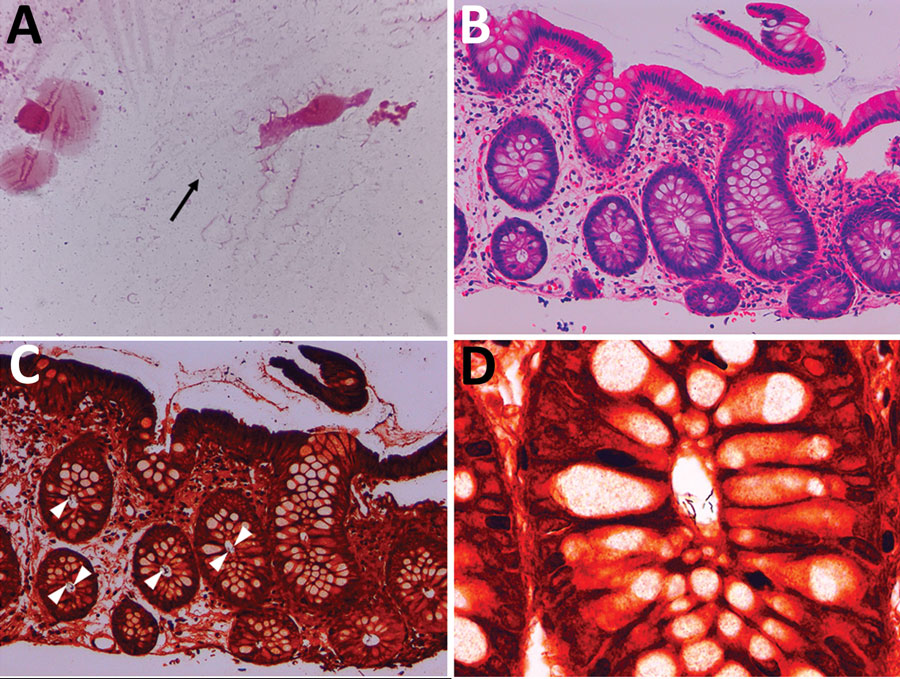
Figure 2. Microscopic findings in ileocolonic biopsy specimens (second colonoscopy) in a man in Japan who had recurrent Helicobacter fennelliae bacteremia. A) Morphologic features of the bacteria in a cecal tissue suspension with Gram staining (original magnification ×2,000). Arrow indicates gram-negative spiral bacilli. B) Histologic findings in a biopsy specimen taken from the transverse colon with hematoxylin-eosin staining (original magnification ×200). The colonic mucosa shows mild leukocytic infiltration. C) Histologic findings in a biopsy specimen taken from the transverse colon with Warthin-Starry silver staining (original magnification ×200). Bacteria are aggregated in crypts (arrowheads) D) Morphologic features of bacteria obtained from the transverse colon with Warthin-Starry silver staining (original magnification ×1,000). A cluster of spiral bacilli was observed.

Main Article

Page created: November 30, 2023
Page updated: December 20, 2023
Page reviewed: December 20, 2023
The conclusions, findings, and opinions expressed by authors contributing to this journal do not necessarily reflect the official position of the U.S. Department of Health and Human Services, the Public Health Service, the Centers for Disease Control and Prevention, or the authors' affiliated institutions. Use of trade names is for identification only and does not imply endorsement by any of the groups named above.
file_external