Enterocytozoon bieneusi Infection after Hematopoietic Stem Cell Transplant in Child, Argentina
Cristian Javier Mena
1, Magalí Pérez Garófalo
1, Juliana Perazzo, Carolina Epelbaum, Gonzalo Castro, Paola Sicilia, Andrés Barnes, Lorena Guasconi, Verónica L. Burstein, Ignacio Beccacece, Mariel A. Almeida, Laura Cervi, Monica Santin, and Laura S. Chiapello

Author affiliations: National University of Córdoba Faculty of Chemical Sciences, Center for Research in Clinical Biochemistry and Immunology, Córdoba, Argentina (C.J. Mena, L. Guasconi, V.L. Burstein, I. Beccacece, M.A. Almeida, L. Cervi, L.S. Chiapello); Dr. Juan P. Garrahan Pediatric Hospital, Buenos Aires, Argentina (M. Pérez Garófalo, J. Perazzo, C. Epelbaum); Central Laboratory of the Province of Córdoba, Córdoba (G. Castro, P. Sicilia); Microbiology Laboratory, Complementary Diagnostic Area, Hospital Rawson, Córdoba (A. Barnes); USDA Agricultural Research Service, Beltsville, Maryland, USA (M. Santin)
Main Article
Figure
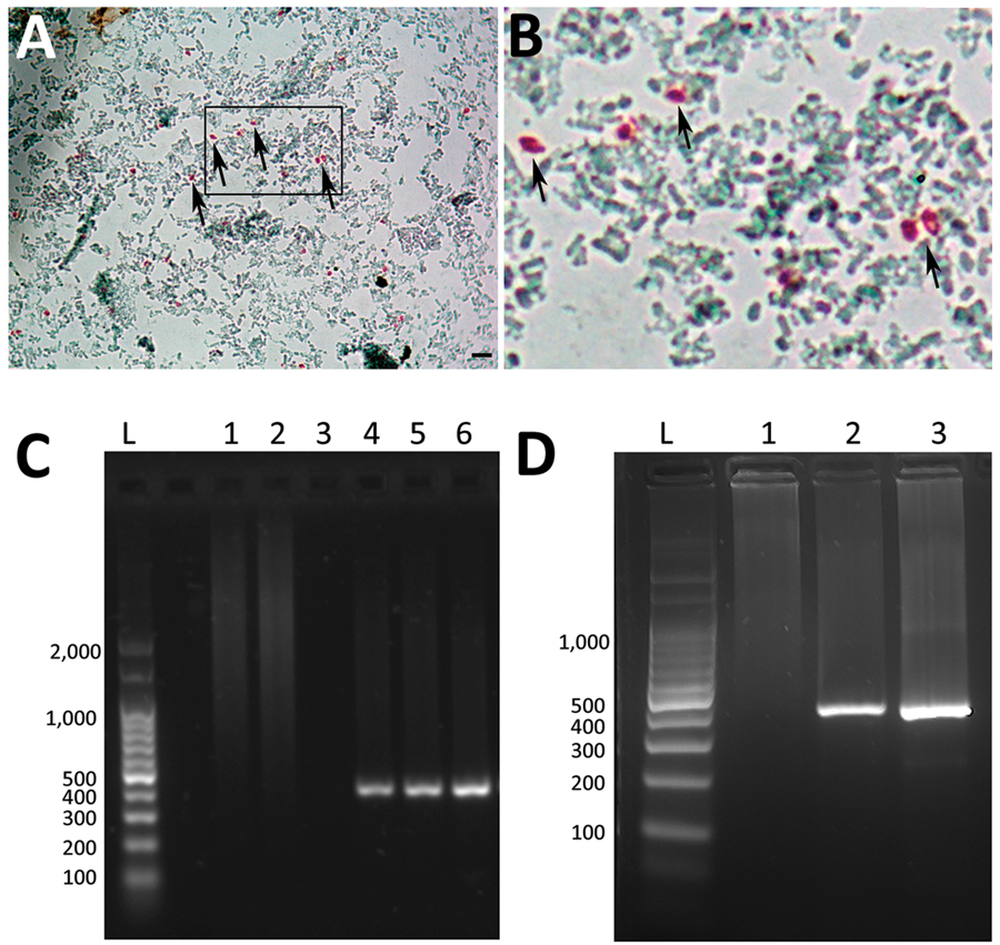
Figure. Enterocytozoon bieneusi detection in fecal sample and liver aspiration biopsy sample from a child with hematopoietic stem cell transplant, Argentina. A) Light microscopy of fecal samples after Weber’s modified trichome staining showing ovoid shaped-spores with a pinkish-red stained wall (arrows). Original magnification × 1,000; scale bars = 5 µm. B) Closer view of boxed area in panel A, showing spores (arrows). C) Agarose gel electrophoresis (2%) showing amplification products (≈390 pb) from nested PCR with inner primers EBITS1 and EBITS2 from patient fecal sample. Lane L, molecular weight ladder; lane 1, negative control (water); lanes 2 and 3, fecal samples from healthy donors; lane 4, positive feces control for E. bieneusi; lanes 5 and 6, fecal samples from patient. D) Nested PCR products from liver aspiration biopsy sample. Lane L, molecular weight ladder; lane 1, fecal sample from healthy donor; lane 2, fecal sample from patient; lane 3, liver aspiration biopsy sample from patient.
Main Article
Page created: January 18, 2024
Page updated: February 22, 2024
Page reviewed: February 22, 2024
The conclusions, findings, and opinions expressed by authors contributing to this journal do not necessarily reflect the official position of the U.S. Department of Health and Human Services, the Public Health Service, the Centers for Disease Control and Prevention, or the authors' affiliated institutions. Use of trade names is for identification only and does not imply endorsement by any of the groups named above.