Synopses
Characteristics and Factors Associated with Death among Patients Hospitalized for Severe Fever with Thrombocytopenia Syndrome, South Korea, 2013 [PDF - 460 KB - 7 pages]
In South Korea, nationwide surveillance for severe fever with thrombocytopenia syndrome (SFTS) began during 2013. Among 301 surveillance cases, 35 hospitalized case-patients in 25 areas were confirmed by using virologic testing, and 16 (46%) case-patients subsequently died. The SFTS cases occurred during May–November and peaked during June (9 cases, 26%). The incidence of SFTS was higher in the southern regions of South Korea. Age and neurologic symptoms, including decreased level of consciousness and slurred speech, were heavily associated with death; neurologic symptoms during the first week after disease onset were also associated with death. Although melena was common among patients who died, no other hemorrhagic manifestations were substantively more common among those who died. No effective treatments, including ribavirin, were identified. Expansion of SFTS surveillance to include the outpatient sector and development of an antibody test would enhance completeness of SFTS detection in South Korea.
| EID | Shin J, Kwon D, Youn S, Park J. Characteristics and Factors Associated with Death among Patients Hospitalized for Severe Fever with Thrombocytopenia Syndrome, South Korea, 2013. Emerg Infect Dis. 2015;21(10):1704-1710. https://doi.org/10.3201/eid2110.141928 |
|---|---|
| AMA | Shin J, Kwon D, Youn S, et al. Characteristics and Factors Associated with Death among Patients Hospitalized for Severe Fever with Thrombocytopenia Syndrome, South Korea, 2013. Emerging Infectious Diseases. 2015;21(10):1704-1710. doi:10.3201/eid2110.141928. |
| APA | Shin, J., Kwon, D., Youn, S., & Park, J. (2015). Characteristics and Factors Associated with Death among Patients Hospitalized for Severe Fever with Thrombocytopenia Syndrome, South Korea, 2013. Emerging Infectious Diseases, 21(10), 1704-1710. https://doi.org/10.3201/eid2110.141928. |
Pacific Broad Tapeworm Adenocephalus pacificus as a Causative Agent of Globally Reemerging Diphyllobothriosis [PDF - 558 KB - 7 pages]
The Pacific broad tapeworm Adenocephalus pacificus (syn. Diphyllobothrium pacificum) is the causative agent of the third most common fish-borne cestodosis among humans. Although most of the nearly 1,000 cases among humans have been reported in South America (Peru, Chile, and Ecuador), cases recently imported to Europe demonstrate the potential for spread of this tapeworm throughout the world as a result of global trade of fresh or chilled marine fish and travel or migration of humans. We provide a comprehensive survey of human cases of infection with this zoonotic parasite, summarize the history of this re-emerging disease, and identify marine fish species that may serve as a source of human infection when eaten raw or undercooked.
| EID | Kuchta R, Serrano-Martínez M, Scholz T. Pacific Broad Tapeworm Adenocephalus pacificus as a Causative Agent of Globally Reemerging Diphyllobothriosis. Emerg Infect Dis. 2015;21(10):1697-1703. https://doi.org/10.3201/eid2110.150516 |
|---|---|
| AMA | Kuchta R, Serrano-Martínez M, Scholz T. Pacific Broad Tapeworm Adenocephalus pacificus as a Causative Agent of Globally Reemerging Diphyllobothriosis. Emerging Infectious Diseases. 2015;21(10):1697-1703. doi:10.3201/eid2110.150516. |
| APA | Kuchta, R., Serrano-Martínez, M., & Scholz, T. (2015). Pacific Broad Tapeworm Adenocephalus pacificus as a Causative Agent of Globally Reemerging Diphyllobothriosis. Emerging Infectious Diseases, 21(10), 1697-1703. https://doi.org/10.3201/eid2110.150516. |
Electronic Public Health Registry of Extensively Drug-Resistant Organisms, Illinois, USA [PDF - 519 KB - 8 pages]
In response to clusters of carbapenem-resistant Enterobacteriaceae (CRE) in Illinois, USA, the Illinois Department of Public Health and the Centers for Disease Control and Prevention Chicago Prevention Epicenter launched a statewide Web-based registry designed for bidirectional data exchange among health care facilities. CRE occurrences are entered and searchable in the system, enabling interfacility communication of patient information. For rapid notification of facilities, admission feeds are automated. During the first 12 months of implementation (November 1, 2013–October 31, 2014), 1,557 CRE reports (≈4.3/day) were submitted from 115 acute care hospitals, 5 long-term acute care hospitals, 46 long-term care facilities, and 7 reference laboratories. Guided by a state and local public health task force of infection prevention specialists and microbiologists and a nonprofit informatics entity, Illinois Department of Public Health deployed a statewide registry of extensively drug-resistant organisms. The legal, technical, and collaborative underpinnings of the system enable rapid incorporation of other emerging organisms.
| EID | Trick WE, Lin MY, Cheng-Leidig R, Driscoll M, Tang AS, Gao W, et al. Electronic Public Health Registry of Extensively Drug-Resistant Organisms, Illinois, USA. Emerg Infect Dis. 2015;21(10):1725-1732. https://doi.org/10.3201/eid2110.150538 |
|---|---|
| AMA | Trick WE, Lin MY, Cheng-Leidig R, et al. Electronic Public Health Registry of Extensively Drug-Resistant Organisms, Illinois, USA. Emerging Infectious Diseases. 2015;21(10):1725-1732. doi:10.3201/eid2110.150538. |
| APA | Trick, W. E., Lin, M. Y., Cheng-Leidig, R., Driscoll, M., Tang, A. S., Gao, W....Weinstein, R. A. (2015). Electronic Public Health Registry of Extensively Drug-Resistant Organisms, Illinois, USA. Emerging Infectious Diseases, 21(10), 1725-1732. https://doi.org/10.3201/eid2110.150538. |
The incidence of severe Haemophilus influenza infections, such as sepsis and meningitis, has declined substantially since the introduction of the H. influenzae serotype b vaccine. However, the H. influenzae type b vaccine fails to protect against nontypeable H. influenzae strains, which have become increasingly frequent causes of invasive disease, especially among children and the elderly. We summarize recent literature supporting the emergence of invasive nontypeable H. influenzae and describe mechanisms that may explain its increasing prevalence over the past 2 decades.
| EID | Langereis JD, de Jonge MI. Invasive Disease Caused by Nontypeable Haemophilus influenzae. Emerg Infect Dis. 2015;21(10):1711-1718. https://doi.org/10.3201/eid2110.150004 |
|---|---|
| AMA | Langereis JD, de Jonge MI. Invasive Disease Caused by Nontypeable Haemophilus influenzae. Emerging Infectious Diseases. 2015;21(10):1711-1718. doi:10.3201/eid2110.150004. |
| APA | Langereis, J. D., & de Jonge, M. I. (2015). Invasive Disease Caused by Nontypeable Haemophilus influenzae. Emerging Infectious Diseases, 21(10), 1711-1718. https://doi.org/10.3201/eid2110.150004. |
We investigated an unusual cluster of 6 patients with Cryptococcus neoformans infection at a community hospital in Arkansas during April–December 2013, to determine source of infection. Four patients had bloodstream infection and 2 had respiratory infection; 3 infections occurred within a 10-day period. Five patients had been admitted to the intensive care unit (ICU) with diagnoses other than cryptococcosis; none had HIV infection, and 1 patient had a history of organ transplantation. We then conducted a retrospective cohort study of all patients admitted to the ICU during April–December 2013 to determine risk factors for cryptococcosis. Four patients with C. neoformans infection had received a short course of steroids; this short-term use was associated with increased risk for cryptococcosis (rate ratio 19.1; 95% CI 2.1–170.0; p<0.01). Although long-term use of steroids is a known risk factor for cryptococcosis, the relationship between short-term steroid use and disease warrants further study
| EID | Vallabhaneni S, Haselow D, Lloyd S, Lockhart SR, Moulton-Meissner H, Lester L, et al. Cluster of Cryptococcus neoformans Infections in Intensive Care Unit, Arkansas, USA, 2013. Emerg Infect Dis. 2015;21(10):1719-1724. https://doi.org/10.3201/eid2110.150249 |
|---|---|
| AMA | Vallabhaneni S, Haselow D, Lloyd S, et al. Cluster of Cryptococcus neoformans Infections in Intensive Care Unit, Arkansas, USA, 2013. Emerging Infectious Diseases. 2015;21(10):1719-1724. doi:10.3201/eid2110.150249. |
| APA | Vallabhaneni, S., Haselow, D., Lloyd, S., Lockhart, S. R., Moulton-Meissner, H., Lester, L....Harris, J. R. (2015). Cluster of Cryptococcus neoformans Infections in Intensive Care Unit, Arkansas, USA, 2013. Emerging Infectious Diseases, 21(10), 1719-1724. https://doi.org/10.3201/eid2110.150249. |
Research
Evolutionary and Ecological Characterization of Mayaro Virus Strains Isolated during an Outbreak, Venezuela, 2010 [PDF - 622 KB - 9 pages]
In 2010, an outbreak of febrile illness with arthralgic manifestations was detected at La Estación village, Portuguesa State, Venezuela. The etiologic agent was determined to be Mayaro virus (MAYV), a reemerging South American alphavirus. A total of 77 cases was reported and 19 were confirmed as seropositive. MAYV was isolated from acute-phase serum samples from 6 symptomatic patients. We sequenced 27 complete genomes representing the full spectrum of MAYV genetic diversity, which facilitated detection of a new genotype, designated N. Phylogenetic analysis of genomic sequences indicated that etiologic strains from Venezuela belong to genotype D. Results indicate that MAYV is highly conserved genetically, showing ≈17% nucleotide divergence across all 3 genotypes and 4% among genotype D strains in the most variable genes. Coalescent analyses suggested genotypes D and L diverged ≈150 years ago and genotype diverged N ≈250 years ago. This virus commonly infects persons residing near enzootic transmission foci because of anthropogenic incursions.
| EID | Auguste AJ, Liria J, Forrester NL, Giambalvo D, Moncada M, Long KC, et al. Evolutionary and Ecological Characterization of Mayaro Virus Strains Isolated during an Outbreak, Venezuela, 2010. Emerg Infect Dis. 2015;21(10):1742-1750. https://doi.org/10.3201/eid2110.141660 |
|---|---|
| AMA | Auguste AJ, Liria J, Forrester NL, et al. Evolutionary and Ecological Characterization of Mayaro Virus Strains Isolated during an Outbreak, Venezuela, 2010. Emerging Infectious Diseases. 2015;21(10):1742-1750. doi:10.3201/eid2110.141660. |
| APA | Auguste, A. J., Liria, J., Forrester, N. L., Giambalvo, D., Moncada, M., Long, K. C....Vasilakis, N. (2015). Evolutionary and Ecological Characterization of Mayaro Virus Strains Isolated during an Outbreak, Venezuela, 2010. Emerging Infectious Diseases, 21(10), 1742-1750. https://doi.org/10.3201/eid2110.141660. |
Delayed Disease Progression in Cynomolgus Macaques Infected with Ebola Virus Makona Strain [PDF - 773 KB - 7 pages]
In late 2013, the largest documented outbreak of Ebola hemorrhagic fever started in Guinea and has since spread to neighboring countries, resulting in almost 27,000 cases and >11,000 deaths in humans. In March 2014, Ebola virus (EBOV) was identified as the causative agent. This study compares the pathogenesis of a new EBOV strain, Makona, which was isolated in Guinea in 2014 with the prototype strain from the 1976 EBOV outbreak in the former Zaire. Both strains cause lethal disease in cynomolgus macaques with similar pathologic changes and hallmark features of Ebola hemorrhagic fever. However, disease progression was delayed in EBOV-Makona–infected animals, suggesting decreased rather than increased virulence of this most recent EBOV strain.
| EID | Marzi A, Feldmann F, Hanley PW, Scott D, Günther S, Feldmann H. Delayed Disease Progression in Cynomolgus Macaques Infected with Ebola Virus Makona Strain. Emerg Infect Dis. 2015;21(10):1777-1783. https://doi.org/10.3201/eid2110.150259 |
|---|---|
| AMA | Marzi A, Feldmann F, Hanley PW, et al. Delayed Disease Progression in Cynomolgus Macaques Infected with Ebola Virus Makona Strain. Emerging Infectious Diseases. 2015;21(10):1777-1783. doi:10.3201/eid2110.150259. |
| APA | Marzi, A., Feldmann, F., Hanley, P. W., Scott, D., Günther, S., & Feldmann, H. (2015). Delayed Disease Progression in Cynomolgus Macaques Infected with Ebola Virus Makona Strain. Emerging Infectious Diseases, 21(10), 1777-1783. https://doi.org/10.3201/eid2110.150259. |
Decreased Ebola Transmission after Rapid Response to Outbreaks in Remote Areas, Liberia, 2014 [PDF - 538 KB - 8 pages]
We measured the reproduction number before and after interventions were implemented to reduce Ebola transmission in 9 outbreaks in Liberia during 2014. We evaluated risk factors for secondary cases and the association between patient admission to an Ebola treatment unit (ETU) and survival. The reproduction number declined 94% from 1.7 (95% CI 1.1–2.6) to 0.1 (95% CI 0.02–0.6) after interventions began. The risk for secondary infections was 90% lower for patients admitted to an ETU (risk ratio 0.1, 95% CI 0.04–0.3) than for those who died in the community. The case-fatality rate was 68% (95% CI 60–74), and ETU admission was associated with a 50% reduction in death (hazard ratio 0.5, 95% CI 0.4–0.8). Isolation and treatment of Ebola patients had the dual benefit of interrupting community transmission and improving survival.
| EID | Lindblade KA, Kateh F, Nagbe TK, Neatherlin JC, Pillai SK, Attfield KR, et al. Decreased Ebola Transmission after Rapid Response to Outbreaks in Remote Areas, Liberia, 2014. Emerg Infect Dis. 2015;21(10):1800-1807. https://doi.org/10.3201/eid2110.150912 |
|---|---|
| AMA | Lindblade KA, Kateh F, Nagbe TK, et al. Decreased Ebola Transmission after Rapid Response to Outbreaks in Remote Areas, Liberia, 2014. Emerging Infectious Diseases. 2015;21(10):1800-1807. doi:10.3201/eid2110.150912. |
| APA | Lindblade, K. A., Kateh, F., Nagbe, T. K., Neatherlin, J. C., Pillai, S. K., Attfield, K. R....Nyenswah, T. G. (2015). Decreased Ebola Transmission after Rapid Response to Outbreaks in Remote Areas, Liberia, 2014. Emerging Infectious Diseases, 21(10), 1800-1807. https://doi.org/10.3201/eid2110.150912. |
Possible Role of Rickettsia felis in Acute Febrile Illness among Children in Gabon [PDF - 706 KB - 8 pages]
Rickettsia felis has been reported to be a cause of fever in sub-Saharan Africa, but this association has been poorly evaluated in Gabon. We assessed the prevalence of this bacterium among children <15 years of age in 4 areas of Gabon; the locations were in urban, semiurban, and rural areas. DNA samples from 410 febrile children and 60 afebrile children were analyzed by quantitative PCR. Overall, the prevalence of R. felis among febrile and afebrile children was 10.2% (42/410 children) and 3.3% (2/60 children), respectively. Prevalence differed among febrile children living in areas that are urban (Franceville, 1.3% [1/77]), semiurban (Koulamoutou, 2.1% [3/141]), and rural (Lastourville, 11.2% [15/134]; Fougamou, 39.7% [23/58]). Furthermore, in a rural area (Fougamou), R. felis was significantly more prevalent in febrile (39.7% [23/58]) than afebrile children (5.0% [1/20]). Additional studies are needed to better understand the pathogenic role of R. felis in this part of the world.
| EID | Mourembou G, Lekana-Douki J, Mediannikov O, Nzondo S, Kouna L, Essone J, et al. Possible Role of Rickettsia felis in Acute Febrile Illness among Children in Gabon. Emerg Infect Dis. 2015;21(10):1808-1815. https://doi.org/10.3201/eid2110.141825 |
|---|---|
| AMA | Mourembou G, Lekana-Douki J, Mediannikov O, et al. Possible Role of Rickettsia felis in Acute Febrile Illness among Children in Gabon. Emerging Infectious Diseases. 2015;21(10):1808-1815. doi:10.3201/eid2110.141825. |
| APA | Mourembou, G., Lekana-Douki, J., Mediannikov, O., Nzondo, S., Kouna, L., Essone, J....Raoult, D. (2015). Possible Role of Rickettsia felis in Acute Febrile Illness among Children in Gabon. Emerging Infectious Diseases, 21(10), 1808-1815. https://doi.org/10.3201/eid2110.141825. |
Induction of Multidrug Tolerance in Plasmodium falciparum by Extended Artemisinin Pressure [PDF - 519 KB - 9 pages]
Plasmodium falciparum resistance to artemisinin derivatives in Southeast Asia threatens global malaria control strategies. Whether delayed parasite clearance, which exposes larger parasite numbers to artemisinins for longer times, selects higher-grade resistance remains unexplored. We investigated whether long-lasting artemisinin pressure selects a novel multidrug-tolerance profile. Although 50% inhibitory concentrations for 10 antimalarial drugs tested were unchanged, drug-tolerant parasites showed higher recrudescence rates for endoperoxides, quinolones, and an antifolate, including partner drugs of recommended combination therapies, but remained susceptible to atovaquone. Moreover, the age range of intraerythrocytic stages able to resist artemisinin was extended to older ring forms and trophozoites. Multidrug tolerance results from drug-induced quiescence, which enables parasites to survive exposure to unrelated antimalarial drugs that inhibit a variety of metabolic pathways. This novel resistance pattern should be urgently monitored in the field because this pattern is not detected by current assays and represents a major threat to antimalarial drug policy.
| EID | Ménard S, Ben Haddou T, Ramadani A, Ariey F, Iriart X, Beghain J, et al. Induction of Multidrug Tolerance in Plasmodium falciparum by Extended Artemisinin Pressure. Emerg Infect Dis. 2015;21(10):1733-1741. https://doi.org/10.3201/eid2110.150682 |
|---|---|
| AMA | Ménard S, Ben Haddou T, Ramadani A, et al. Induction of Multidrug Tolerance in Plasmodium falciparum by Extended Artemisinin Pressure. Emerging Infectious Diseases. 2015;21(10):1733-1741. doi:10.3201/eid2110.150682. |
| APA | Ménard, S., Ben Haddou, T., Ramadani, A., Ariey, F., Iriart, X., Beghain, J....Benoit-Vical, F. (2015). Induction of Multidrug Tolerance in Plasmodium falciparum by Extended Artemisinin Pressure. Emerging Infectious Diseases, 21(10), 1733-1741. https://doi.org/10.3201/eid2110.150682. |
Epidemiology of Lyme Disease, Nova Scotia, Canada, 2002–2013 [PDF - 533 KB - 8 pages]
Ixodes scapularis ticks, which transmit Borrelia burgdorferi, the causative agent of Lyme disease (LD), are endemic to at least 6 regions of Nova Scotia, Canada. To assess the epidemiology and prevalence of LD in Nova Scotia, we analyzed data from 329 persons with LD reported in Nova Scotia during 2002–2013. Most patients reported symptoms of early localized infection with rash (89.7%), influenza-like illness (69.6%), or both; clinician-diagnosed erythema migrans was documented for 53.2%. In a separate serosurvey, of 1,855 serum samples screened for antibodies to B. burgdorferi, 2 were borderline positive (both with an indeterminate IgG on Western blot), resulting in an estimated seroprevalence of 0.14% (95% CI 0.02%–0.51%). Although LD incidence in Nova Scotia has risen sharply since 2002 and is the highest in Canada (16/100,000 population in 2013), the estimated number of residents with evidence of infection is low, and risk is localized to currently identified LD-endemic regions.
| EID | Hatchette TF, Johnston B, Schleihauf E, Mask A, Haldane D, Drebot M, et al. Epidemiology of Lyme Disease, Nova Scotia, Canada, 2002–2013. Emerg Infect Dis. 2015;21(10):1751-1758. https://doi.org/10.3201/eid2110.141640 |
|---|---|
| AMA | Hatchette TF, Johnston B, Schleihauf E, et al. Epidemiology of Lyme Disease, Nova Scotia, Canada, 2002–2013. Emerging Infectious Diseases. 2015;21(10):1751-1758. doi:10.3201/eid2110.141640. |
| APA | Hatchette, T. F., Johnston, B., Schleihauf, E., Mask, A., Haldane, D., Drebot, M....Lindsay, R. (2015). Epidemiology of Lyme Disease, Nova Scotia, Canada, 2002–2013. Emerging Infectious Diseases, 21(10), 1751-1758. https://doi.org/10.3201/eid2110.141640. |
Haemaphysalis longicornis Ticks as Reservoir and Vector of Severe Fever with Thrombocytopenia Syndrome Virus in China [PDF - 583 KB - 7 pages]
Severe fever with thrombocytopenia syndrome (SFTS) is an emerging hemorrhagic fever in East Asia caused by SFTS virus (SFTSV), a newly discovered phlebovirus. The Haemaphysalis longicornis tick has been suspected to be the vector of SFTSV. To determine whether SFTSV can be transmitted among ticks, from ticks to animals, and from animals to ticks, we conducted transmission studies between developmental stages of H. longicornis ticks and between ticks and mice. Using reverse transcription PCR, we also analyzed the prevalence of SFTSV infection among H. longicornis ticks collected from vegetation in Shandong Province, China. Our results showed a low prevalence of SFTSV among collected ticks (0.2%, 8/3,300 ticks), and we showed that ticks fed on SFTSV-infected mice could acquire the virus and transstadially and transovarially transmit it to other developmental stages of ticks. Furthermore, SFTSV-infected ticks could transmit the virus to mice during feeding. Our findings indicate ticks could serve as a vector and reservoir of SFTSV.
| EID | Luo L, Zhao L, Wen H, Zhang Z, Liu J, Fang L, et al. Haemaphysalis longicornis Ticks as Reservoir and Vector of Severe Fever with Thrombocytopenia Syndrome Virus in China. Emerg Infect Dis. 2015;21(10):1770-1776. https://doi.org/10.3201/eid2110.150126 |
|---|---|
| AMA | Luo L, Zhao L, Wen H, et al. Haemaphysalis longicornis Ticks as Reservoir and Vector of Severe Fever with Thrombocytopenia Syndrome Virus in China. Emerging Infectious Diseases. 2015;21(10):1770-1776. doi:10.3201/eid2110.150126. |
| APA | Luo, L., Zhao, L., Wen, H., Zhang, Z., Liu, J., Fang, L....Yu, X. (2015). Haemaphysalis longicornis Ticks as Reservoir and Vector of Severe Fever with Thrombocytopenia Syndrome Virus in China. Emerging Infectious Diseases, 21(10), 1770-1776. https://doi.org/10.3201/eid2110.150126. |
Effect of Live Poultry Market Closure on Avian Influenza A(H7N9) Virus Activity in Guangzhou, China, 2014 [PDF - 611 KB - 10 pages]
We assessed the effect of closing live poultry markets in China on influenza A(H7N9) virus detection and viability. Intensive sampling was carried out before, during, and after a 2-week citywide market closure; the markets were cleaned and disinfected at the beginning of the closure period. Swab samples were collected at different sites within the markets and tested for H7N9 by real-time reverse transcription PCR and culture. During the closure, H7N9 viral RNA detection and isolation rates in retail markets decreased by 79% (95% CI 64%–88%) and 92% (95% CI 58%–98%), respectively. However, viable H7N9 virus could be cultured from wastewater samples collected up to 2 days after the market closure began. Our findings indicates that poultry workers and the general population are constantly exposed to H7N9 virus at these markets and that market closure and disinfection rapidly reduces the amount of viable virus.
| EID | Yuan J, Lau E, Li K, Leung Y, Yang Z, Xie C, et al. Effect of Live Poultry Market Closure on Avian Influenza A(H7N9) Virus Activity in Guangzhou, China, 2014. Emerg Infect Dis. 2015;21(10):1784-1793. https://doi.org/10.3201/eid2110.150623 |
|---|---|
| AMA | Yuan J, Lau E, Li K, et al. Effect of Live Poultry Market Closure on Avian Influenza A(H7N9) Virus Activity in Guangzhou, China, 2014. Emerging Infectious Diseases. 2015;21(10):1784-1793. doi:10.3201/eid2110.150623. |
| APA | Yuan, J., Lau, E., Li, K., Leung, Y., Yang, Z., Xie, C....Peiris, M. (2015). Effect of Live Poultry Market Closure on Avian Influenza A(H7N9) Virus Activity in Guangzhou, China, 2014. Emerging Infectious Diseases, 21(10), 1784-1793. https://doi.org/10.3201/eid2110.150623. |
Environmental Factors Related to Fungal Wound Contamination after Combat Trauma in Afghanistan, 2009–2011 [PDF - 658 KB - 11 pages]
During the recent war in Afghanistan (2001–2014), invasive fungal wound infections (IFIs) among US combat casualties were associated with risk factors related to the mechanism and pattern of injury. Although previous studies recognized that IFI patients primarily sustained injuries in southern Afghanistan, environmental data were not examined. We compared environmental conditions of this region with those of an area in eastern Afghanistan that was not associated with observed IFIs after injury. A larger proportion of personnel injured in the south (61%) grew mold from wound cultures than those injured in the east (20%). In a multivariable analysis, the southern location, characterized by lower elevation, warmer temperatures, and greater isothermality, was independently associated with mold contamination of wounds. These environmental characteristics, along with known risk factors related to injury characteristics, may be useful in modeling the risk for IFIs after traumatic injury in other regions.
| EID | Tribble DR, Rodriguez CJ, Weintrob AC, Shaikh F, Aggarwal D, Carson M, et al. Environmental Factors Related to Fungal Wound Contamination after Combat Trauma in Afghanistan, 2009–2011. Emerg Infect Dis. 2015;21(10):1759-1769. https://doi.org/10.3201/eid2110.141759 |
|---|---|
| AMA | Tribble DR, Rodriguez CJ, Weintrob AC, et al. Environmental Factors Related to Fungal Wound Contamination after Combat Trauma in Afghanistan, 2009–2011. Emerging Infectious Diseases. 2015;21(10):1759-1769. doi:10.3201/eid2110.141759. |
| APA | Tribble, D. R., Rodriguez, C. J., Weintrob, A. C., Shaikh, F., Aggarwal, D., Carson, M....Masuoka, P. (2015). Environmental Factors Related to Fungal Wound Contamination after Combat Trauma in Afghanistan, 2009–2011. Emerging Infectious Diseases, 21(10), 1759-1769. https://doi.org/10.3201/eid2110.141759. |
Human Infection with Ehrlichia muris–like Pathogen, United States, 2007–2013 [PDF - 449 KB - 6 pages]
An Ehrlichia muris–like (EML) pathogen was detected among 4 patients in Minnesota and Wisconsin during 2009. We characterized additional cases clinically and epidemiologically. During 2004–2013, blood samples from 75,077 patients from all 50 United States were tested by PCR from the groEL gene for Ehrlichia spp. and Anaplasma phagocytophilum. During 2007–2013, samples from 69 (0.1%) patients were positive for the EML pathogen; patients were from 5 states: Indiana (1), Michigan (1), Minnesota (33), North Dakota (3), and Wisconsin (31). Most (64%) patients were male; median age was 63 (range 15–94) years; and all 69 patients reported likely tick exposure in Minnesota or Wisconsin. Fever, malaise, thrombocytopenia, and lymphopenia were the most common symptoms. Sixteen (23%) patients were hospitalized (median 4 days); all recovered, and 96% received doxycycline. Infection with the EML pathogen should be considered for persons reporting tick exposure in Minnesota or Wisconsin.
| EID | Johnson DK, Schiffman E, Davis JP, Neitzel D, Sloan LM, Nicholson WL, et al. Human Infection with Ehrlichia muris–like Pathogen, United States, 2007–2013. Emerg Infect Dis. 2015;21(10):1794-1799. https://doi.org/10.3201/eid2110.150143 |
|---|---|
| AMA | Johnson DK, Schiffman E, Davis JP, et al. Human Infection with Ehrlichia muris–like Pathogen, United States, 2007–2013. Emerging Infectious Diseases. 2015;21(10):1794-1799. doi:10.3201/eid2110.150143. |
| APA | Johnson, D. K., Schiffman, E., Davis, J. P., Neitzel, D., Sloan, L. M., Nicholson, W. L....Pritt, B. (2015). Human Infection with Ehrlichia muris–like Pathogen, United States, 2007–2013. Emerging Infectious Diseases, 21(10), 1794-1799. https://doi.org/10.3201/eid2110.150143. |
Dispatches
Methicillin-Susceptible, Vancomycin-Resistant Staphylococcus aureus, Brazil [PDF - 553 KB - 5 pages]
We report characterization of a methicillin-susceptible, vancomycin-resistant bloodstream isolate of Staphylococcus aureus recovered from a patient in Brazil. Emergence of vancomycin resistance in methicillin-susceptible S. aureus would indicate that this resistance trait might be poised to disseminate more rapidly among S. aureus and represents a major public health threat.
| EID | Panesso D, Planet P, Diaz L, Hugonnet J, Tran TT, Narechania A, et al. Methicillin-Susceptible, Vancomycin-Resistant Staphylococcus aureus, Brazil. Emerg Infect Dis. 2015;21(10):1844-1848. https://doi.org/10.3201/eid2110.141914 |
|---|---|
| AMA | Panesso D, Planet P, Diaz L, et al. Methicillin-Susceptible, Vancomycin-Resistant Staphylococcus aureus, Brazil. Emerging Infectious Diseases. 2015;21(10):1844-1848. doi:10.3201/eid2110.141914. |
| APA | Panesso, D., Planet, P., Diaz, L., Hugonnet, J., Tran, T. T., Narechania, A....Arias, C. A. (2015). Methicillin-Susceptible, Vancomycin-Resistant Staphylococcus aureus, Brazil. Emerging Infectious Diseases, 21(10), 1844-1848. https://doi.org/10.3201/eid2110.141914. |
Public Health Response to Aedes aegypti and Ae. albopictus Mosquitoes Invading California, USA [PDF - 401 KB - 3 pages]
Aedes aegypti and Ae. albopictus mosquitoes, primary vectors of dengue and chikungunya viruses, were recently detected in California, USA. The threat of potential local transmission of these viruses increases as more infected travelers arrive from affected areas. Public health response has included enhanced human and mosquito surveillance, education, and intensive mosquito control.
| EID | Porse C, Kramer VL, Yoshimizu M, Metzger ME, Hu R, Padgett K, et al. Public Health Response to Aedes aegypti and Ae. albopictus Mosquitoes Invading California, USA. Emerg Infect Dis. 2015;21(10):1827-1829. https://doi.org/10.3201/eid2110.150494 |
|---|---|
| AMA | Porse C, Kramer VL, Yoshimizu M, et al. Public Health Response to Aedes aegypti and Ae. albopictus Mosquitoes Invading California, USA. Emerging Infectious Diseases. 2015;21(10):1827-1829. doi:10.3201/eid2110.150494. |
| APA | Porse, C., Kramer, V. L., Yoshimizu, M., Metzger, M. E., Hu, R., Padgett, K....Vugia, D. (2015). Public Health Response to Aedes aegypti and Ae. albopictus Mosquitoes Invading California, USA. Emerging Infectious Diseases, 21(10), 1827-1829. https://doi.org/10.3201/eid2110.150494. |
Acute Flaccid Paralysis Associated with Novel Enterovirus C105 [PDF - 377 KB - 3 pages]
An outbreak of acute flaccid paralysis among children in the United States during summer 2014 was tentatively associated with enterovirus D68 infection. This syndrome in a child in fall 2014 was associated with enterovirus C105 infection. The presence of this virus strain in North America may pose a diagnostic challenge.
| EID | Horner LM, Poulter MD, Brenton J, Turner RB. Acute Flaccid Paralysis Associated with Novel Enterovirus C105. Emerg Infect Dis. 2015;21(10):1858-1860. https://doi.org/10.3201/eid2110.150759 |
|---|---|
| AMA | Horner LM, Poulter MD, Brenton J, et al. Acute Flaccid Paralysis Associated with Novel Enterovirus C105. Emerging Infectious Diseases. 2015;21(10):1858-1860. doi:10.3201/eid2110.150759. |
| APA | Horner, L. M., Poulter, M. D., Brenton, J., & Turner, R. B. (2015). Acute Flaccid Paralysis Associated with Novel Enterovirus C105. Emerging Infectious Diseases, 21(10), 1858-1860. https://doi.org/10.3201/eid2110.150759. |
Utility of Oral Swab Sampling for Ebola Virus Detection in Guinea Pig Model [PDF - 433 KB - 4 pages]
To determine the utility of oral swabs for diagnosing infection with Ebola virus, we used a guinea pig model and obtained daily antemortem and postmortem swab samples. According to quantitative reverse transcription PCR analysis, the diagnostic value was poor for antemortem swab samples but excellent for postmortem samples.
| EID | Spengler JR, Chakrabarti AK, Coleman-McCray JD, Martin BE, Nichol ST, Spiropoulou CF, et al. Utility of Oral Swab Sampling for Ebola Virus Detection in Guinea Pig Model. Emerg Infect Dis. 2015;21(10):1816-1819. https://doi.org/10.3201/eid2110.150840 |
|---|---|
| AMA | Spengler JR, Chakrabarti AK, Coleman-McCray JD, et al. Utility of Oral Swab Sampling for Ebola Virus Detection in Guinea Pig Model. Emerging Infectious Diseases. 2015;21(10):1816-1819. doi:10.3201/eid2110.150840. |
| APA | Spengler, J. R., Chakrabarti, A. K., Coleman-McCray, J. D., Martin, B. E., Nichol, S. T., Spiropoulou, C. F....Bird, B. H. (2015). Utility of Oral Swab Sampling for Ebola Virus Detection in Guinea Pig Model. Emerging Infectious Diseases, 21(10), 1816-1819. https://doi.org/10.3201/eid2110.150840. |
Local and International Implications of Schistosomiasis Acquired in Corsica, France [PDF - 522 KB - 4 pages]
We report 11 cases of schistosomiasis in international travelers who had bathed in rivers in Corsica, France, during 2012–2014. The infections were diagnosed in 2014 and reported to the GeoSentinel Surveillance Network and European Travel Medicine Network. Travelers can be sentinels for emerging infections; thus, this situation warrants a concerted human and veterinary epidemiologic response.
| EID | Gautret P, Mockenhaupt FP, von Sonnenburg F, Rothe C, Libman M, Van De Winkel K, et al. Local and International Implications of Schistosomiasis Acquired in Corsica, France. Emerg Infect Dis. 2015;21(10):1865-1868. https://doi.org/10.3201/eid2110.150881 |
|---|---|
| AMA | Gautret P, Mockenhaupt FP, von Sonnenburg F, et al. Local and International Implications of Schistosomiasis Acquired in Corsica, France. Emerging Infectious Diseases. 2015;21(10):1865-1868. doi:10.3201/eid2110.150881. |
| APA | Gautret, P., Mockenhaupt, F. P., von Sonnenburg, F., Rothe, C., Libman, M., Van De Winkel, K....Schlagenhauf, P. (2015). Local and International Implications of Schistosomiasis Acquired in Corsica, France. Emerging Infectious Diseases, 21(10), 1865-1868. https://doi.org/10.3201/eid2110.150881. |
Detection of Mixed Infections with Plasmodium spp. by PCR, India, 2014 [PDF - 528 KB - 5 pages]
In 8 malaria-endemic states in India, mixed Plasmodium spp. infections were detected by PCR in 17.4% (265/1,521) of blood samples that microscopy had shown to contain only P. falciparum. The quality of microscopy must be improved because use of PCR for detection of malaria parasites is limited in rural areas.
| EID | Krishna S, Bharti PK, Chandel HS, Ahmad A, Kumar R, Singh PP, et al. Detection of Mixed Infections with Plasmodium spp. by PCR, India, 2014. Emerg Infect Dis. 2015;21(10):1853-1857. https://doi.org/10.3201/eid2110.150678 |
|---|---|
| AMA | Krishna S, Bharti PK, Chandel HS, et al. Detection of Mixed Infections with Plasmodium spp. by PCR, India, 2014. Emerging Infectious Diseases. 2015;21(10):1853-1857. doi:10.3201/eid2110.150678. |
| APA | Krishna, S., Bharti, P. K., Chandel, H. S., Ahmad, A., Kumar, R., Singh, P. P....Singh, N. (2015). Detection of Mixed Infections with Plasmodium spp. by PCR, India, 2014. Emerging Infectious Diseases, 21(10), 1853-1857. https://doi.org/10.3201/eid2110.150678. |
Taeniasis among Refugees Living on Thailand–Myanmar Border, 2012 [PDF - 432 KB - 3 pages]
We tested refugee camp residents on the Thailand–Myanmar border for Taenia solium infection. Taeniasis prevalence was consistent with that for other disease-endemic regions, but seropositivity indicating T. solium taeniasis was rare. Seropositivity indicating cysticercosis was 5.5% in humans, and 3.2% in pigs. Corralling pigs and providing latrines may control transmission of these tapeworms within this camp.
| EID | McCleery EJ, Patchanee P, Pongsopawijit P, Chailangkarn S, Tiwananthagorn S, Jongchansittoe P, et al. Taeniasis among Refugees Living on Thailand–Myanmar Border, 2012. Emerg Infect Dis. 2015;21(10):1824-1826. https://doi.org/10.3201/eid2110.141657 |
|---|---|
| AMA | McCleery EJ, Patchanee P, Pongsopawijit P, et al. Taeniasis among Refugees Living on Thailand–Myanmar Border, 2012. Emerging Infectious Diseases. 2015;21(10):1824-1826. doi:10.3201/eid2110.141657. |
| APA | McCleery, E. J., Patchanee, P., Pongsopawijit, P., Chailangkarn, S., Tiwananthagorn, S., Jongchansittoe, P....O’Neal, S. E. (2015). Taeniasis among Refugees Living on Thailand–Myanmar Border, 2012. Emerging Infectious Diseases, 21(10), 1824-1826. https://doi.org/10.3201/eid2110.141657. |
Influenza Virus Surveillance in Coordinated Swine Production Systems, United States [PDF - 446 KB - 3 pages]
To clarify the epidemiology of influenza A viruses in coordinated swine production systems to which no animals from outside the system are introduced, we conducted virologic surveillance during September 2012–September 2013. Animal age, geographic location, and farm type were found to affect the prevalence of these viruses.
| EID | Kaplan B, DeBeauchamp J, Stigger-Rosser E, Franks J, Crumpton J, Turner J, et al. Influenza Virus Surveillance in Coordinated Swine Production Systems, United States. Emerg Infect Dis. 2015;21(10):1834-1836. https://doi.org/10.3201/eid2110.140633 |
|---|---|
| AMA | Kaplan B, DeBeauchamp J, Stigger-Rosser E, et al. Influenza Virus Surveillance in Coordinated Swine Production Systems, United States. Emerging Infectious Diseases. 2015;21(10):1834-1836. doi:10.3201/eid2110.140633. |
| APA | Kaplan, B., DeBeauchamp, J., Stigger-Rosser, E., Franks, J., Crumpton, J., Turner, J....Lowe, J. F. (2015). Influenza Virus Surveillance in Coordinated Swine Production Systems, United States. Emerging Infectious Diseases, 21(10), 1834-1836. https://doi.org/10.3201/eid2110.140633. |
Heartland Virus Neutralizing Antibodies in Vertebrate Wildlife, United States, 2009–2014 [PDF - 423 KB - 4 pages]
Since its discovery in 2009, the tickborne Heartland virus (HRTV) has caused human illness in Missouri, Oklahoma, and Tennessee USA. To better assess the geographic distribution of HRTV, we used wildlife serology as an indicator. This retrospective evaluation determined that HRTV is widespread within the central and eastern United States.
| EID | Riemersma KK, Komar N. Heartland Virus Neutralizing Antibodies in Vertebrate Wildlife, United States, 2009–2014. Emerg Infect Dis. 2015;21(10):1830-1833. https://doi.org/10.3201/eid2110.150380 |
|---|---|
| AMA | Riemersma KK, Komar N. Heartland Virus Neutralizing Antibodies in Vertebrate Wildlife, United States, 2009–2014. Emerging Infectious Diseases. 2015;21(10):1830-1833. doi:10.3201/eid2110.150380. |
| APA | Riemersma, K. K., & Komar, N. (2015). Heartland Virus Neutralizing Antibodies in Vertebrate Wildlife, United States, 2009–2014. Emerging Infectious Diseases, 21(10), 1830-1833. https://doi.org/10.3201/eid2110.150380. |
Spatiotemporal Patterns of Schistosomiasis-Related Deaths, Brazil, 2000–2011 [PDF - 670 KB - 4 pages]
We analyzed spatiotemporal patterns of 8,756 schistosomiasis-related deaths in Brazil during 2000–2011 and identified high-risk clusters of deaths, mainly in highly schistosomiasis-endemic areas along the coast of Brazil’s Northeast Region. Schistosomiasis remains a neglected public health problem with a high number of deaths in disease-endemic and emerging focal areas.
| EID | Martins-Melo F, Pinheiro M, Ramos A, Alencar C, Bezerra F, Heukelbach J. Spatiotemporal Patterns of Schistosomiasis-Related Deaths, Brazil, 2000–2011. Emerg Infect Dis. 2015;21(10):1820-1823. https://doi.org/10.3201/eid2110.141438 |
|---|---|
| AMA | Martins-Melo F, Pinheiro M, Ramos A, et al. Spatiotemporal Patterns of Schistosomiasis-Related Deaths, Brazil, 2000–2011. Emerging Infectious Diseases. 2015;21(10):1820-1823. doi:10.3201/eid2110.141438. |
| APA | Martins-Melo, F., Pinheiro, M., Ramos, A., Alencar, C., Bezerra, F., & Heukelbach, J. (2015). Spatiotemporal Patterns of Schistosomiasis-Related Deaths, Brazil, 2000–2011. Emerging Infectious Diseases, 21(10), 1820-1823. https://doi.org/10.3201/eid2110.141438. |
Cross-sectional Serosurvey of Crimean-Congo Hemorrhagic Fever Virus IgG in Livestock, India, 2013–2014 [PDF - 306 KB - 3 pages]
We conducted a cross-sectional serosurvey of Crimean-Congo hemorrhagic fever (CCHF) among livestock in 22 states and 1 union territory of India. A total of 5,636 samples from bovines, sheep, and goats were screened for CCHF virus IgG. IgG was detected in 354 samples, indicating that this virus is widespread in this country.
| EID | Mourya DT, Yadav PD, Shete AM, Sathe PS, Sarkale P, Pattnaik B, et al. Cross-sectional Serosurvey of Crimean-Congo Hemorrhagic Fever Virus IgG in Livestock, India, 2013–2014. Emerg Infect Dis. 2015;21(10):1837-1839. https://doi.org/10.3201/eid2110.141961 |
|---|---|
| AMA | Mourya DT, Yadav PD, Shete AM, et al. Cross-sectional Serosurvey of Crimean-Congo Hemorrhagic Fever Virus IgG in Livestock, India, 2013–2014. Emerging Infectious Diseases. 2015;21(10):1837-1839. doi:10.3201/eid2110.141961. |
| APA | Mourya, D. T., Yadav, P. D., Shete, A. M., Sathe, P. S., Sarkale, P., Pattnaik, B....Katoch, V. M. (2015). Cross-sectional Serosurvey of Crimean-Congo Hemorrhagic Fever Virus IgG in Livestock, India, 2013–2014. Emerging Infectious Diseases, 21(10), 1837-1839. https://doi.org/10.3201/eid2110.141961. |
Risk Factors for Sustained Cholera Transmission, Juba County, South Sudan, 2014 [PDF - 392 KB - 4 pages]
We conducted a case–control study to identify risk factors for the 2014 cholera outbreak in Juba County, South Sudan. Illness was associated with traveling or eating away from home; treating drinking water and receiving oral cholera vaccination were protective. Oral cholera vaccination should be used to complement cholera prevention efforts.
| EID | Ujjiga T, Wamala JF, Mogga J, Othwonh TO, Mutonga D, Kone-Coulibaly A, et al. Risk Factors for Sustained Cholera Transmission, Juba County, South Sudan, 2014. Emerg Infect Dis. 2015;21(10):1849-1852. https://doi.org/10.3201/eid2110.142051 |
|---|---|
| AMA | Ujjiga T, Wamala JF, Mogga J, et al. Risk Factors for Sustained Cholera Transmission, Juba County, South Sudan, 2014. Emerging Infectious Diseases. 2015;21(10):1849-1852. doi:10.3201/eid2110.142051. |
| APA | Ujjiga, T., Wamala, J. F., Mogga, J., Othwonh, T. O., Mutonga, D., Kone-Coulibaly, A....Rumunu, J. (2015). Risk Factors for Sustained Cholera Transmission, Juba County, South Sudan, 2014. Emerging Infectious Diseases, 21(10), 1849-1852. https://doi.org/10.3201/eid2110.142051. |
Transmission Risk from Imported Plasmodium vivax Malaria in the China–Myanmar Border Region [PDF - 437 KB - 4 pages]
Malaria importation and local vector susceptibility to imported Plasmodium vivax infection are a continuing risk along the China–Myanmar border. Malaria transmission has been prevented in 3 border villages in Tengchong County, Yunnan Province, China, by use of active fever surveillance, integrated vector control measures, and intensified surveillance and response.
| EID | Wang D, Li S, Cheng Z, Xiao N, Cotter C, Hwang J, et al. Transmission Risk from Imported Plasmodium vivax Malaria in the China–Myanmar Border Region. Emerg Infect Dis. 2015;21(10):1861-1864. https://doi.org/10.3201/eid2110.150679 |
|---|---|
| AMA | Wang D, Li S, Cheng Z, et al. Transmission Risk from Imported Plasmodium vivax Malaria in the China–Myanmar Border Region. Emerging Infectious Diseases. 2015;21(10):1861-1864. doi:10.3201/eid2110.150679. |
| APA | Wang, D., Li, S., Cheng, Z., Xiao, N., Cotter, C., Hwang, J....Wang, S. (2015). Transmission Risk from Imported Plasmodium vivax Malaria in the China–Myanmar Border Region. Emerging Infectious Diseases, 21(10), 1861-1864. https://doi.org/10.3201/eid2110.150679. |
Novel Paramyxoviruses in Bats from Sub-Saharan Africa, 2007–2012 [PDF - 418 KB - 4 pages]
As part of a larger survey for detection of pathogens among wildlife in sub-Saharan Africa conducted during 2007–2012, multiple diverse paramyxovirus sequences were detected in renal tissues of bats. Phylogenetic analysis supports the presence of at least 2 major viral lineages and suggests that paramyxoviruses are strongly associated with several bat genera.
| EID | Mortlock M, Kuzmin IV, Weyer J, Gilbert AT, Agwanda B, Rupprecht CE, et al. Novel Paramyxoviruses in Bats from Sub-Saharan Africa, 2007–2012. Emerg Infect Dis. 2015;21(10):1840-1843. https://doi.org/10.3201/eid2110.140368 |
|---|---|
| AMA | Mortlock M, Kuzmin IV, Weyer J, et al. Novel Paramyxoviruses in Bats from Sub-Saharan Africa, 2007–2012. Emerging Infectious Diseases. 2015;21(10):1840-1843. doi:10.3201/eid2110.140368. |
| APA | Mortlock, M., Kuzmin, I. V., Weyer, J., Gilbert, A. T., Agwanda, B., Rupprecht, C. E....Markotter, W. (2015). Novel Paramyxoviruses in Bats from Sub-Saharan Africa, 2007–2012. Emerging Infectious Diseases, 21(10), 1840-1843. https://doi.org/10.3201/eid2110.140368. |
Letters
Zika Virus Outbreak, Bahia, Brazil [PDF - 270 KB - 2 pages]
| EID | Campos GS, Bandeira AC, Sardi SI. Zika Virus Outbreak, Bahia, Brazil. Emerg Infect Dis. 2015;21(10):1885-1886. https://doi.org/10.3201/eid2110.150847 |
|---|---|
| AMA | Campos GS, Bandeira AC, Sardi SI. Zika Virus Outbreak, Bahia, Brazil. Emerging Infectious Diseases. 2015;21(10):1885-1886. doi:10.3201/eid2110.150847. |
| APA | Campos, G. S., Bandeira, A. C., & Sardi, S. I. (2015). Zika Virus Outbreak, Bahia, Brazil. Emerging Infectious Diseases, 21(10), 1885-1886. https://doi.org/10.3201/eid2110.150847. |
Bloody Diarrhea Associated with Hookworm Infection in Traveler Returning to France from Myanmar [PDF - 318 KB - 2 pages]
| EID | Brunet J, Lemoine J, Lefebvre N, Denis J, Pfaff AW, Abou-Bacar A, et al. Bloody Diarrhea Associated with Hookworm Infection in Traveler Returning to France from Myanmar. Emerg Infect Dis. 2015;21(10):1878-1879. https://doi.org/10.3201/eid2110.150695 |
|---|---|
| AMA | Brunet J, Lemoine J, Lefebvre N, et al. Bloody Diarrhea Associated with Hookworm Infection in Traveler Returning to France from Myanmar. Emerging Infectious Diseases. 2015;21(10):1878-1879. doi:10.3201/eid2110.150695. |
| APA | Brunet, J., Lemoine, J., Lefebvre, N., Denis, J., Pfaff, A. W., Abou-Bacar, A....Candolfi, E. (2015). Bloody Diarrhea Associated with Hookworm Infection in Traveler Returning to France from Myanmar. Emerging Infectious Diseases, 21(10), 1878-1879. https://doi.org/10.3201/eid2110.150695. |
Schistosomiasis Screening of Travelers from Italy with Possible Exposure in Corsica, France [PDF - 432 KB - 3 pages]
| EID | Beltrame A, Zammarchi L, Zuglian G, Gobbi F, Angheben A, Marchese V, et al. Schistosomiasis Screening of Travelers from Italy with Possible Exposure in Corsica, France. Emerg Infect Dis. 2015;21(10):1887-1889. https://doi.org/10.3201/eid2110.150869 |
|---|---|
| AMA | Beltrame A, Zammarchi L, Zuglian G, et al. Schistosomiasis Screening of Travelers from Italy with Possible Exposure in Corsica, France. Emerging Infectious Diseases. 2015;21(10):1887-1889. doi:10.3201/eid2110.150869. |
| APA | Beltrame, A., Zammarchi, L., Zuglian, G., Gobbi, F., Angheben, A., Marchese, V....Bisoffi, Z. (2015). Schistosomiasis Screening of Travelers from Italy with Possible Exposure in Corsica, France. Emerging Infectious Diseases, 21(10), 1887-1889. https://doi.org/10.3201/eid2110.150869. |
Transmission of Entamoeba nuttalli and Trichuris trichiura from Nonhuman Primates to Humans [PDF - 272 KB - 2 pages]
| EID | Levecke B, Dorny P, Vercammen F, Visser LG, Van Esbroeck M, Vercruysse J, et al. Transmission of Entamoeba nuttalli and Trichuris trichiura from Nonhuman Primates to Humans. Emerg Infect Dis. 2015;21(10):1871-1872. https://doi.org/10.3201/eid2110.141456 |
|---|---|
| AMA | Levecke B, Dorny P, Vercammen F, et al. Transmission of Entamoeba nuttalli and Trichuris trichiura from Nonhuman Primates to Humans. Emerging Infectious Diseases. 2015;21(10):1871-1872. doi:10.3201/eid2110.141456. |
| APA | Levecke, B., Dorny, P., Vercammen, F., Visser, L. G., Van Esbroeck, M., Vercruysse, J....Verweij, J. J. (2015). Transmission of Entamoeba nuttalli and Trichuris trichiura from Nonhuman Primates to Humans. Emerging Infectious Diseases, 21(10), 1871-1872. https://doi.org/10.3201/eid2110.141456. |
Human Infections with Pseudoterranova cattani Nematodes, Chile [PDF - 342 KB - 2 pages]
| EID | Weitzel T, Sugiyama H, Yamasaki H, Ramirez C, Rosas R, Mercado R. Human Infections with Pseudoterranova cattani Nematodes, Chile. Emerg Infect Dis. 2015;21(10):1874-1875. https://doi.org/10.3201/eid2110.141848 |
|---|---|
| AMA | Weitzel T, Sugiyama H, Yamasaki H, et al. Human Infections with Pseudoterranova cattani Nematodes, Chile. Emerging Infectious Diseases. 2015;21(10):1874-1875. doi:10.3201/eid2110.141848. |
| APA | Weitzel, T., Sugiyama, H., Yamasaki, H., Ramirez, C., Rosas, R., & Mercado, R. (2015). Human Infections with Pseudoterranova cattani Nematodes, Chile. Emerging Infectious Diseases, 21(10), 1874-1875. https://doi.org/10.3201/eid2110.141848. |
Association of Necrotizing Wounds Colonized by Maggots with Ignatzschineria–Associated Septicemia [PDF - 392 KB - 3 pages]
| EID | Le Brun C, Gombert M, Robert S, Mercier E, Lanotte P. Association of Necrotizing Wounds Colonized by Maggots with Ignatzschineria–Associated Septicemia. Emerg Infect Dis. 2015;21(10):1881-1883. https://doi.org/10.3201/eid2110.150748 |
|---|---|
| AMA | Le Brun C, Gombert M, Robert S, et al. Association of Necrotizing Wounds Colonized by Maggots with Ignatzschineria–Associated Septicemia. Emerging Infectious Diseases. 2015;21(10):1881-1883. doi:10.3201/eid2110.150748. |
| APA | Le Brun, C., Gombert, M., Robert, S., Mercier, E., & Lanotte, P. (2015). Association of Necrotizing Wounds Colonized by Maggots with Ignatzschineria–Associated Septicemia. Emerging Infectious Diseases, 21(10), 1881-1883. https://doi.org/10.3201/eid2110.150748. |
Alaria alata Mesocercariae among Feral Cats and Badgers, Denmark [PDF - 334 KB - 3 pages]
| EID | Takeuchi-Storm N, Al-Sabi M, Thamsborg SM, Enemark HL. Alaria alata Mesocercariae among Feral Cats and Badgers, Denmark. Emerg Infect Dis. 2015;21(10):1872-1874. https://doi.org/10.3201/eid2110.141817 |
|---|---|
| AMA | Takeuchi-Storm N, Al-Sabi M, Thamsborg SM, et al. Alaria alata Mesocercariae among Feral Cats and Badgers, Denmark. Emerging Infectious Diseases. 2015;21(10):1872-1874. doi:10.3201/eid2110.141817. |
| APA | Takeuchi-Storm, N., Al-Sabi, M., Thamsborg, S. M., & Enemark, H. L. (2015). Alaria alata Mesocercariae among Feral Cats and Badgers, Denmark. Emerging Infectious Diseases, 21(10), 1872-1874. https://doi.org/10.3201/eid2110.141817. |
Burkholderia pseudomallei Infection in US Traveler Returning from Mexico, 2014 [PDF - 271 KB - 2 pages]
| EID | Cheng JW, Hayden MK, Singh K, Heimler I, Gee JE, Proia L, et al. Burkholderia pseudomallei Infection in US Traveler Returning from Mexico, 2014. Emerg Infect Dis. 2015;21(10):1884-1885. https://doi.org/10.3201/eid2110.150815 |
|---|---|
| AMA | Cheng JW, Hayden MK, Singh K, et al. Burkholderia pseudomallei Infection in US Traveler Returning from Mexico, 2014. Emerging Infectious Diseases. 2015;21(10):1884-1885. doi:10.3201/eid2110.150815. |
| APA | Cheng, J. W., Hayden, M. K., Singh, K., Heimler, I., Gee, J. E., Proia, L....Sha, B. E. (2015). Burkholderia pseudomallei Infection in US Traveler Returning from Mexico, 2014. Emerging Infectious Diseases, 21(10), 1884-1885. https://doi.org/10.3201/eid2110.150815. |
Familial Trichostrongylus Infection Misdiagnosed as Acute Fascioliasis [PDF - 328 KB - 2 pages]
| EID | Ashrafi K, Tahbaz A, Sharifdini M, Mas-Coma S. Familial Trichostrongylus Infection Misdiagnosed as Acute Fascioliasis. Emerg Infect Dis. 2015;21(10):1869-1870. https://doi.org/10.3201/eid2110.141392 |
|---|---|
| AMA | Ashrafi K, Tahbaz A, Sharifdini M, et al. Familial Trichostrongylus Infection Misdiagnosed as Acute Fascioliasis. Emerging Infectious Diseases. 2015;21(10):1869-1870. doi:10.3201/eid2110.141392. |
| APA | Ashrafi, K., Tahbaz, A., Sharifdini, M., & Mas-Coma, S. (2015). Familial Trichostrongylus Infection Misdiagnosed as Acute Fascioliasis. Emerging Infectious Diseases, 21(10), 1869-1870. https://doi.org/10.3201/eid2110.141392. |
Severe Sepsis Caused by California Serogroup Orthobunyavirus [PDF - 303 KB - 2 pages]
| EID | Rogstad DK, Schiffman E, Neitzel D, Baddour LM. Severe Sepsis Caused by California Serogroup Orthobunyavirus. Emerg Infect Dis. 2015;21(10):1876-1877. https://doi.org/10.3201/eid2110.150394 |
|---|---|
| AMA | Rogstad DK, Schiffman E, Neitzel D, et al. Severe Sepsis Caused by California Serogroup Orthobunyavirus. Emerging Infectious Diseases. 2015;21(10):1876-1877. doi:10.3201/eid2110.150394. |
| APA | Rogstad, D. K., Schiffman, E., Neitzel, D., & Baddour, L. M. (2015). Severe Sepsis Caused by California Serogroup Orthobunyavirus. Emerging Infectious Diseases, 21(10), 1876-1877. https://doi.org/10.3201/eid2110.150394. |
Vaccine-Derived Polioviruses Not Detected by Global Surveillance Screening Assay [PDF - 271 KB - 2 pages]
| EID | Sharma DK, Nalavade UP, Varose SY, Deshpande JM. Vaccine-Derived Polioviruses Not Detected by Global Surveillance Screening Assay. Emerg Infect Dis. 2015;21(10):1880-1881. https://doi.org/10.3201/eid2110.150702 |
|---|---|
| AMA | Sharma DK, Nalavade UP, Varose SY, et al. Vaccine-Derived Polioviruses Not Detected by Global Surveillance Screening Assay. Emerging Infectious Diseases. 2015;21(10):1880-1881. doi:10.3201/eid2110.150702. |
| APA | Sharma, D. K., Nalavade, U. P., Varose, S. Y., & Deshpande, J. M. (2015). Vaccine-Derived Polioviruses Not Detected by Global Surveillance Screening Assay. Emerging Infectious Diseases, 21(10), 1880-1881. https://doi.org/10.3201/eid2110.150702. |
Zika Virus Transmission from French Polynesia to Brazil [PDF - 591 KB - 1 page]
| EID | Musso D. Zika Virus Transmission from French Polynesia to Brazil. Emerg Infect Dis. 2015;21(10):1887. https://doi.org/10.3201/eid2110.151125 |
|---|---|
| AMA | Musso D. Zika Virus Transmission from French Polynesia to Brazil. Emerging Infectious Diseases. 2015;21(10):1887. doi:10.3201/eid2110.151125. |
| APA | Musso, D. (2015). Zika Virus Transmission from French Polynesia to Brazil. Emerging Infectious Diseases, 21(10), 1887. https://doi.org/10.3201/eid2110.151125. |
Books and Media
Anthropology of Infectious Disease [PDF - 218 KB - 1 page]
| EID | Munyikwa M. Anthropology of Infectious Disease. Emerg Infect Dis. 2015;21(10):1890. https://doi.org/10.3201/eid2110.151263 |
|---|---|
| AMA | Munyikwa M. Anthropology of Infectious Disease. Emerging Infectious Diseases. 2015;21(10):1890. doi:10.3201/eid2110.151263. |
| APA | Munyikwa, M. (2015). Anthropology of Infectious Disease. Emerging Infectious Diseases, 21(10), 1890. https://doi.org/10.3201/eid2110.151263. |
Etymologia
Etymologia: Ignatzschineria [PDF - 300 KB - 1 page]
| EID | Etymologia: Ignatzschineria. Emerg Infect Dis. 2015;21(10):1883. https://doi.org/10.3201/eid2110.et2110 |
|---|---|
| AMA | Etymologia: Ignatzschineria. Emerging Infectious Diseases. 2015;21(10):1883. doi:10.3201/eid2110.et2110. |
| APA | (2015). Etymologia: Ignatzschineria. Emerging Infectious Diseases, 21(10), 1883. https://doi.org/10.3201/eid2110.et2110. |
Corrections
Correction: Vol. 21, No. 7 [PDF - 262 KB - 1 page]
| EID | Correction: Vol. 21, No. 7. Emerg Infect Dis. 2015;21(10):1877. https://doi.org/10.3201/eid2110.c12110 |
|---|---|
| AMA | Correction: Vol. 21, No. 7. Emerging Infectious Diseases. 2015;21(10):1877. doi:10.3201/eid2110.c12110. |
| APA | (2015). Correction: Vol. 21, No. 7. Emerging Infectious Diseases, 21(10), 1877. https://doi.org/10.3201/eid2110.c12110. |
About the Cover
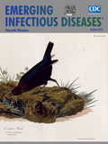
Don’t Lay Your Eggs All in One Basket: Brood Parasitism as a Survival Strategy [PDF - 305 KB - 2 pages]
| EID | Breedlove B, Arguin PM. Don’t Lay Your Eggs All in One Basket: Brood Parasitism as a Survival Strategy. Emerg Infect Dis. 2015;21(10):1891-1892. https://doi.org/10.3201/eid2110.ac2110 |
|---|---|
| AMA | Breedlove B, Arguin PM. Don’t Lay Your Eggs All in One Basket: Brood Parasitism as a Survival Strategy. Emerging Infectious Diseases. 2015;21(10):1891-1892. doi:10.3201/eid2110.ac2110. |
| APA | Breedlove, B., & Arguin, P. M. (2015). Don’t Lay Your Eggs All in One Basket: Brood Parasitism as a Survival Strategy. Emerging Infectious Diseases, 21(10), 1891-1892. https://doi.org/10.3201/eid2110.ac2110. |