Synopses
Outbreak of HIV Infection Linked to Nosocomial Transmission, China, 2016–2017 [PDF - 2.07 MB - 9 pages]
On January 25, 2017, a physician from ZC Hospital in Hangzhou, China, reported to the Zhejiang Provincial Center for Disease Control and Prevention that a potential HIV outbreak might have occurred during lymphocyte immunotherapy (LIT) performed at the hospital on December 30, 2016. We immediately began investigating and identified the index case-patient as an LIT patient’s husband who donated lymphocytes for his wife’s LIT and later screened HIV-reactive. Subsequent contamination by a technician resulted in the potential exposure of 34 LIT patients. Acute HIV infection was diagnosed in 5 persons. Phylogenetic analysis confirmed that the HIV-1 gag, pol, and env gene sequences from the index and outbreak-related cases had >99.5% similarity. Rapid investigation and implementation of effective control measures successfully controlled the outbreak. This incident provides evidence of a lapse in infection control causing HIV transmission, highlighting the need for stronger measures to protect patients from infectious disease exposure.
| EID | Pan X, Jiang J, Ma Q, Zhang J, Yang J, Chen W, et al. Outbreak of HIV Infection Linked to Nosocomial Transmission, China, 2016–2017. Emerg Infect Dis. 2018;24(12):2141-2149. https://doi.org/10.3201/eid2412.180117 |
|---|---|
| AMA | Pan X, Jiang J, Ma Q, et al. Outbreak of HIV Infection Linked to Nosocomial Transmission, China, 2016–2017. Emerging Infectious Diseases. 2018;24(12):2141-2149. doi:10.3201/eid2412.180117. |
| APA | Pan, X., Jiang, J., Ma, Q., Zhang, J., Yang, J., Chen, W....Wu, Z. (2018). Outbreak of HIV Infection Linked to Nosocomial Transmission, China, 2016–2017. Emerging Infectious Diseases, 24(12), 2141-2149. https://doi.org/10.3201/eid2412.180117. |
We summarize and analyze historical and current data regarding the reemergence of St. Louis encephalitis virus (SLEV; genus Flavivirus) in the Americas. Historically, SLEV caused encephalitis outbreaks in the United States; however, it was not considered a public health concern in the rest of the Americas. After the introduction of West Nile virus in 1999, activity of SLEV decreased considerably in the United States. During 2014–2015, SLEV caused a human outbreak in Arizona and caused isolated human cases in California in 2016 and 2017. Phylogenetic analyses indicate that the emerging SLEV in the western United States is related to the epidemic strains isolated during a human encephalitis outbreak in Córdoba, Argentina, in 2005. Ecoepidemiologic studies suggest that the emergence of SLEV in Argentina was caused by the introduction of a more pathogenic strain and increasing populations of the eared dove (amplifying host).
| EID | Diaz A, Coffey LL, Burkett-Cadena N, Day JF. Reemergence of St. Louis Encephalitis Virus in the Americas. Emerg Infect Dis. 2018;24(12):2150-2157. https://doi.org/10.3201/eid2412.180372 |
|---|---|
| AMA | Diaz A, Coffey LL, Burkett-Cadena N, et al. Reemergence of St. Louis Encephalitis Virus in the Americas. Emerging Infectious Diseases. 2018;24(12):2150-2157. doi:10.3201/eid2412.180372. |
| APA | Diaz, A., Coffey, L. L., Burkett-Cadena, N., & Day, J. F. (2018). Reemergence of St. Louis Encephalitis Virus in the Americas. Emerging Infectious Diseases, 24(12), 2150-2157. https://doi.org/10.3201/eid2412.180372. |
Autochthonous Human Case of Seoul Virus Infection, the Netherlands [PDF - 767 KB - 6 pages]
Orthohantaviruses are a group of rodentborne viruses with a worldwide distribution. The orthohantavirus Seoul virus (SEOV) can cause hemorrhagic fever with renal syndrome in humans and is distributed worldwide, like its reservoir host, the rat. Cases of SEOV in wild and pet rats have been described in several countries, and human cases have been reported in the United Kingdom, France, Canada, and the United States. In the Netherlands, SEOV has previously been found in wild brown rats. We describe an autochthonous human case of SEOV infection in the Netherlands. This patient had nonspecific clinical symptoms of an orthohantavirus infection (gastrointestinal symptoms and distinct elevation of liver enzymes). Subsequent source investigation revealed 2 potential sources, the patient’s feeder rats and a feeder rat farm. At both sources, a high prevalence of SEOV was found in the rats. The virus closely resembled the Cherwell and Turckheim SEOV strains that were previously found in Europe.
| EID | Swanink C, Reimerink J, Gisolf J, de Vries A, Claassen M, Martens L, et al. Autochthonous Human Case of Seoul Virus Infection, the Netherlands. Emerg Infect Dis. 2018;24(12):2158-2163. https://doi.org/10.3201/eid2412.180229 |
|---|---|
| AMA | Swanink C, Reimerink J, Gisolf J, et al. Autochthonous Human Case of Seoul Virus Infection, the Netherlands. Emerging Infectious Diseases. 2018;24(12):2158-2163. doi:10.3201/eid2412.180229. |
| APA | Swanink, C., Reimerink, J., Gisolf, J., de Vries, A., Claassen, M., Martens, L....Maas, M. (2018). Autochthonous Human Case of Seoul Virus Infection, the Netherlands. Emerging Infectious Diseases, 24(12), 2158-2163. https://doi.org/10.3201/eid2412.180229. |
Restaurant Inspection Letter Grades and Salmonella Infections, New York, New York, USA [PDF - 506 KB - 5 pages]
Rates of Salmonella infection in the United States have not changed over the past 20 years. Restaurants are frequent settings for Salmonella outbreaks and sporadic infections. Few studies have examined the effect of posting letter grades for restaurant inspections on the incidence of foodborne illness. We compared Salmonella infection rates in New York, New York, USA (NYC), with those in the rest of New York state before and after implementation of a letter grade system for restaurant inspections in NYC. We calculated a segmented regression model for interrupted time series data. After implementation of letter grading, the rate of Salmonella infections decreased 5.3% per year in NYC versus the rest of New York state during 2011–2015, compared with the period before implementation, 2006–2010. Posting restaurant inspection results as letter grades at the point of service was associated with a decline in Salmonella infections in NYC and warrants consideration for broader use.
| EID | Firestone MJ, Hedberg CW. Restaurant Inspection Letter Grades and Salmonella Infections, New York, New York, USA. Emerg Infect Dis. 2018;24(12):2164-2168. https://doi.org/10.3201/eid2412.180544 |
|---|---|
| AMA | Firestone MJ, Hedberg CW. Restaurant Inspection Letter Grades and Salmonella Infections, New York, New York, USA. Emerging Infectious Diseases. 2018;24(12):2164-2168. doi:10.3201/eid2412.180544. |
| APA | Firestone, M. J., & Hedberg, C. W. (2018). Restaurant Inspection Letter Grades and Salmonella Infections, New York, New York, USA. Emerging Infectious Diseases, 24(12), 2164-2168. https://doi.org/10.3201/eid2412.180544. |
Research
Spatial Analysis of Wildlife Tuberculosis Based on a Serologic Survey Using Dried Blood Spots, Portugal [PDF - 1.70 MB - 7 pages]
We investigated the spatial epidemiology of bovine tuberculosis (TB) in wildlife in a multihost system. We surveyed bovine TB in Portugal by serologic analysis of elutes of dried blood spots obtained from hunted wild boar. We modeled spatial disease risk by using areal generalized linear mixed models with conditional autoregressive priors. Antibodies against Mycobaterium bovis were detected in 2.4% (95% CI 1.5%–3.8%) of 678 wild boar in 2 geographic clusters, and the predicted risk fits well with independent reports of M. bovis culture. Results show that elutes are an almost perfect substitute for serum (Cohen unweighted κ = 0.818), indicating that serologic tests coupled with dried blood spots are an effective strategy for large-scale bovine TB surveys, using wild boar as sentinel species. Results also show that bovine TB is an emerging wildlife disease and stress the need to prevent further geographic spread and prevalence increase.
| EID | Santos N, Nunes T, Fonseca C, Vieira-Pinto M, Almeida V, Gortázar C, et al. Spatial Analysis of Wildlife Tuberculosis Based on a Serologic Survey Using Dried Blood Spots, Portugal. Emerg Infect Dis. 2018;24(12):2169-2175. https://doi.org/10.3201/eid2412.171357 |
|---|---|
| AMA | Santos N, Nunes T, Fonseca C, et al. Spatial Analysis of Wildlife Tuberculosis Based on a Serologic Survey Using Dried Blood Spots, Portugal. Emerging Infectious Diseases. 2018;24(12):2169-2175. doi:10.3201/eid2412.171357. |
| APA | Santos, N., Nunes, T., Fonseca, C., Vieira-Pinto, M., Almeida, V., Gortázar, C....Correia-Neves, M. (2018). Spatial Analysis of Wildlife Tuberculosis Based on a Serologic Survey Using Dried Blood Spots, Portugal. Emerging Infectious Diseases, 24(12), 2169-2175. https://doi.org/10.3201/eid2412.171357. |
Rat Lungworm Infection in Rodents across Post-Katrina New Orleans, Louisiana, USA [PDF - 2.29 MB - 8 pages]
Rat lungworm (Angiostrongylus cantonensis), a parasitic nematode that can cause eosinophilic meningitis in humans, was first detected in New Orleans, Louisiana, USA, in the mid-1980s and now appears to be widespread in the southeastern United States. We assessed the distribution, prevalence, and intensity of A. cantonensis infection in New Orleans by examining lung biopsy samples of rodents trapped at 96 sites in 9 areas in Orleans Parish and 1 area in neighboring St. Bernard Parish during May 2015 through February 2017. These areas were selected to capture contrasting levels of income, flooding, and pos-disaster landscape management after Hurricane Katrina in 2005. We detected A. cantonensis in all areas and in 3 of the 4 rat species trapped. Overall prevalence was ≈38% but varied by area, host species, and host species co-occurrence. Infection intensity also varied by host species. These findings suggest that socioecological analysis of heterogeneity in definitive and intermediate host infection could improve understanding of health risks across the city.
| EID | Rael RC, Peterson AC, Ghersi-Chavez B, Riegel C, Lesen AE, Blum MJ. Rat Lungworm Infection in Rodents across Post-Katrina New Orleans, Louisiana, USA. Emerg Infect Dis. 2018;24(12):2176-2183. https://doi.org/10.3201/eid2412.180056 |
|---|---|
| AMA | Rael RC, Peterson AC, Ghersi-Chavez B, et al. Rat Lungworm Infection in Rodents across Post-Katrina New Orleans, Louisiana, USA. Emerging Infectious Diseases. 2018;24(12):2176-2183. doi:10.3201/eid2412.180056. |
| APA | Rael, R. C., Peterson, A. C., Ghersi-Chavez, B., Riegel, C., Lesen, A. E., & Blum, M. J. (2018). Rat Lungworm Infection in Rodents across Post-Katrina New Orleans, Louisiana, USA. Emerging Infectious Diseases, 24(12), 2176-2183. https://doi.org/10.3201/eid2412.180056. |
Terrestrial Bird Migration and West Nile Virus Circulation, United States [PDF - 2.70 MB - 11 pages]
Host migration and emerging pathogens are strongly associated, especially with regard to zoonotic diseases. West Nile virus (WNV), a mosquitoborne pathogen capable of causing severe, sometimes fatal, neuroinvasive disease in humans, is maintained in highly mobile avian hosts. Using phylogeographic approaches, we investigated the relationship between WNV circulation in the United States and the flight paths of terrestrial birds. We demonstrated southward migration of WNV in the eastern flyway and northward migration in the central flyway, which is consistent with the looped flight paths of many terrestrial birds. We also identified 3 optimal locations for targeted WNV surveillance campaigns in the United States—Illinois, New York, and Texas. These results illustrate the value of multidisciplinary approaches to surveillance of infectious diseases, especially zoonotic diseases.
| EID | Swetnam D, Widen SG, Wood TG, Reyna M, Wilkerson L, Debboun M, et al. Terrestrial Bird Migration and West Nile Virus Circulation, United States. Emerg Infect Dis. 2018;24(12):2184-2194. https://doi.org/10.3201/eid2412.180382 |
|---|---|
| AMA | Swetnam D, Widen SG, Wood TG, et al. Terrestrial Bird Migration and West Nile Virus Circulation, United States. Emerging Infectious Diseases. 2018;24(12):2184-2194. doi:10.3201/eid2412.180382. |
| APA | Swetnam, D., Widen, S. G., Wood, T. G., Reyna, M., Wilkerson, L., Debboun, M....Barrett, A. (2018). Terrestrial Bird Migration and West Nile Virus Circulation, United States. Emerging Infectious Diseases, 24(12), 2184-2194. https://doi.org/10.3201/eid2412.180382. |
Capnocytophaga canimorsus Capsular Serovar and Disease Severity, Helsinki Hospital District, Finland, 2000–2017 [PDF - 1.86 MB - 7 pages]
We assembled a collection of 73 Capnocytophaga canimorsus isolates obtained from blood cultures taken from patients treated at Helsinki University Hospital (Helsinki, Finland) during 2000–2017. We serotyped these isolates by PCR and Western blot and attempted to correlate pathogen serovar with patient characteristics. Our analyses showed, in agreement with previous research, that 3 C. canimorsus serovars (A–C) caused most (91.8%) human infections, despite constituting only 7.6% of isolates found in dogs. The 3 fatalities that occurred in our cohort were equally represented by these serovars. We found 2 untypeable isolates, which we designated serovars J and K. We did not detect an association between serovar and disease severity, immune status, alcohol abuse, or smoking status, but dog bites occurred more frequently among patients infected with non-A–C serovars. Future research is needed to confirm serovar virulence and develop strategies to reduce risk for these infections in humans.
| EID | Hess E, Renzi F, Karhunen P, Dol M, Lefèvre A, Antikainen J, et al. Capnocytophaga canimorsus Capsular Serovar and Disease Severity, Helsinki Hospital District, Finland, 2000–2017. Emerg Infect Dis. 2018;24(12):2195-2201. https://doi.org/10.3201/eid2412.172060 |
|---|---|
| AMA | Hess E, Renzi F, Karhunen P, et al. Capnocytophaga canimorsus Capsular Serovar and Disease Severity, Helsinki Hospital District, Finland, 2000–2017. Emerging Infectious Diseases. 2018;24(12):2195-2201. doi:10.3201/eid2412.172060. |
| APA | Hess, E., Renzi, F., Karhunen, P., Dol, M., Lefèvre, A., Antikainen, J....Cornelis, G. R. (2018). Capnocytophaga canimorsus Capsular Serovar and Disease Severity, Helsinki Hospital District, Finland, 2000–2017. Emerging Infectious Diseases, 24(12), 2195-2201. https://doi.org/10.3201/eid2412.172060. |
Crimean-Congo Hemorrhagic Fever Virus, Mongolia, 2013–2014 [PDF - 910 KB - 8 pages]
During 2013–2014, we collected 1,926 serum samples from humans and 4,583 ticks (Hyalomma asiaticum or Dermacentor nuttalli) in select regions of Mongolia to determine the risk for Crimean-Congo hemorrhagic fever virus (CCHFV) infection among humans in this country. Testing of human serum samples by ELISA demonstrated an overall CCHFV antibody prevalence of 1.4%; Bayankhongor Province had the highest prevalence, 2.63%. We pooled and analyzed tick specimens by real-time reverse transcription PCR; 1 CCHFV-positive H. asiaticum tick pool from Ömnögovi was identified. In phylogenetic analyses, the virus’s partial small segment clustered with CCHFV isolates from Central Asia, and the complete medium segment grouped with CCHFV isolates from Africa, Asia, and the Middle East. This study confirms CCHFV endemicity in Mongolia and provides information on risk for CCHFV infection. Further research is needed to better define the risk for CCHFV disease to improve risk mitigation, diagnostics, and surveillance.
| EID | Voorhees MA, Padilla SL, Jamsransuren D, Koehler JW, Delp KL, Adiyadorj D, et al. Crimean-Congo Hemorrhagic Fever Virus, Mongolia, 2013–2014. Emerg Infect Dis. 2018;24(12):2202-2209. https://doi.org/10.3201/eid2412.180175 |
|---|---|
| AMA | Voorhees MA, Padilla SL, Jamsransuren D, et al. Crimean-Congo Hemorrhagic Fever Virus, Mongolia, 2013–2014. Emerging Infectious Diseases. 2018;24(12):2202-2209. doi:10.3201/eid2412.180175. |
| APA | Voorhees, M. A., Padilla, S. L., Jamsransuren, D., Koehler, J. W., Delp, K. L., Adiyadorj, D....Schoepp, R. J. (2018). Crimean-Congo Hemorrhagic Fever Virus, Mongolia, 2013–2014. Emerging Infectious Diseases, 24(12), 2202-2209. https://doi.org/10.3201/eid2412.180175. |
Novel Type of Chronic Wasting Disease Detected in Moose (Alces alces), Norway [PDF - 2.31 MB - 9 pages]
Chronic wasting disease (CWD) persists in cervid populations of North America and in 2016 was detected for the first time in Europe in a wild reindeer in Norway. We report the detection of CWD in 3 moose (Alces alces) in Norway, identified through a large scale surveillance program. The cases occurred in 13–14-year-old female moose, and we detected an abnormal form of prion protein (PrPSc) in the brain but not in lymphoid tissues. Immunohistochemistry revealed that the moose shared the same neuropathologic phenotype, characterized by mostly intraneuronal deposition of PrPSc. This pattern differed from that observed in reindeer and has not been previously reported in CWD-infected cervids. Moreover, Western blot revealed a PrPSc type distinguishable from previous CWD cases and from known ruminant prion diseases in Europe, with the possible exception of sheep CH1641. These findings suggest that these cases in moose represent a novel type of CWD.
| EID | Pirisinu L, Tran L, Chiappini B, Vanni I, Di Bari MA, Vaccari G, et al. Novel Type of Chronic Wasting Disease Detected in Moose (Alces alces), Norway. Emerg Infect Dis. 2018;24(12):2210-2218. https://doi.org/10.3201/eid2412.180702 |
|---|---|
| AMA | Pirisinu L, Tran L, Chiappini B, et al. Novel Type of Chronic Wasting Disease Detected in Moose (Alces alces), Norway. Emerging Infectious Diseases. 2018;24(12):2210-2218. doi:10.3201/eid2412.180702. |
| APA | Pirisinu, L., Tran, L., Chiappini, B., Vanni, I., Di Bari, M. A., Vaccari, G....Benestad, S. L. (2018). Novel Type of Chronic Wasting Disease Detected in Moose (Alces alces), Norway. Emerging Infectious Diseases, 24(12), 2210-2218. https://doi.org/10.3201/eid2412.180702. |
Genomic Characterization of β-Glucuronidase–Positive Escherichia coli O157:H7 Producing Stx2a [PDF - 2.97 MB - 9 pages]
Among Shiga toxin (Stx)–producing Escherichia coli (STEC) O157:H7 strains, those producing Stx2a cause more severe diseases. Atypical STEC O157:H7 strains showing a β-glucuronidase–positive phenotype (GP STEC O157:H7) have rarely been isolated from humans, mostly from persons with asymptomatic or mild infections; Stx2a-producing strains have not been reported. We isolated, from a patient with bloody diarrhea, a GP STEC O157:H7 strain (PV15-279) that produces Stx2a in addition to Stx1a and Stx2c. Genomic comparison with other STEC O157 strains revealed that PV15-279 recently emerged from the stx1a/stx2c-positive GP STEC O157:H7 clone circulating in Japan. Major virulence genes are shared between typical (β-glucuronidase–negative) and GP STEC O157:H7 strains, and the Stx2-producing ability of PV15-279 is comparable to that of typical STEC O157:H7 strains; therefore, PV15-279 presents a virulence potential similar to that of typical STEC O157:H7. This study reveals the importance of GP O157:H7 as a source of highly pathogenic STEC clones.
| EID | Ogura Y, Seto K, Morimoto Y, Nakamura K, Sato MP, Gotoh Y, et al. Genomic Characterization of β-Glucuronidase–Positive Escherichia coli O157:H7 Producing Stx2a. Emerg Infect Dis. 2018;24(12):2219-2227. https://doi.org/10.3201/eid2412.180404 |
|---|---|
| AMA | Ogura Y, Seto K, Morimoto Y, et al. Genomic Characterization of β-Glucuronidase–Positive Escherichia coli O157:H7 Producing Stx2a. Emerging Infectious Diseases. 2018;24(12):2219-2227. doi:10.3201/eid2412.180404. |
| APA | Ogura, Y., Seto, K., Morimoto, Y., Nakamura, K., Sato, M. P., Gotoh, Y....Hayashi, T. (2018). Genomic Characterization of β-Glucuronidase–Positive Escherichia coli O157:H7 Producing Stx2a. Emerging Infectious Diseases, 24(12), 2219-2227. https://doi.org/10.3201/eid2412.180404. |
Survey of Ebola Viruses in Frugivorous and Insectivorous Bats in Guinea, Cameroon, and the Democratic Republic of the Congo, 2015–2017
To clarify the role of bats in the ecology of Ebola viruses, we assessed the prevalence of Ebola virus antibodies in a large-scale sample of bats collected during 2015–2017 from countries in Africa that have had previous Ebola outbreaks (Guinea, the Democratic Republic of the Congo) or are at high risk for outbreaks (Cameroon). We analyzed 4,022 blood samples of bats from >12 frugivorous and 27 insectivorous species; 2–37 (0.05%–0.92%) bats were seropositive for Zaire and 0–30 (0%–0.75%) bats for Sudan Ebola viruses. We observed Ebola virus antibodies in 1 insectivorous bat genus and 6 frugivorous bat species. Certain bat species widespread across Africa had serologic evidence of Zaire and Sudan Ebola viruses. No viral RNA was detected in the subset of samples tested (n = 665). Ongoing surveillance of bats and other potential animal reservoirs are required to predict and prepare for future outbreaks.
| EID | De Nys HM, Kingebeni P, Keita AK, Butel C, Thaurignac G, Villabona-Arenas C, et al. Survey of Ebola Viruses in Frugivorous and Insectivorous Bats in Guinea, Cameroon, and the Democratic Republic of the Congo, 2015–2017. Emerg Infect Dis. 2018;24(12):2228-2240. https://doi.org/10.3201/eid2412.180740 |
|---|---|
| AMA | De Nys HM, Kingebeni P, Keita AK, et al. Survey of Ebola Viruses in Frugivorous and Insectivorous Bats in Guinea, Cameroon, and the Democratic Republic of the Congo, 2015–2017. Emerging Infectious Diseases. 2018;24(12):2228-2240. doi:10.3201/eid2412.180740. |
| APA | De Nys, H. M., Kingebeni, P., Keita, A. K., Butel, C., Thaurignac, G., Villabona-Arenas, C....Peeters, M. (2018). Survey of Ebola Viruses in Frugivorous and Insectivorous Bats in Guinea, Cameroon, and the Democratic Republic of the Congo, 2015–2017. Emerging Infectious Diseases, 24(12), 2228-2240. https://doi.org/10.3201/eid2412.180740. |
Rat Hepatitis E Virus as Cause of Persistent Hepatitis after Liver Transplant [PDF - 3.36 MB - 10 pages]
All hepatitis E virus (HEV) variants reported to infect humans belong to the species Orthohepevirus A (HEV-A). The zoonotic potential of the species Orthohepevirus C (HEV-C), which circulates in rats and is highly divergent from HEV-A, is unknown. We report a liver transplant recipient with hepatitis caused by HEV-C infection. We detected HEV-C RNA in multiple clinical samples and HEV-C antigen in the liver. The complete genome of the HEV-C isolate had 93.7% nt similarity to an HEV-C strain from Vietnam. The patient had preexisting HEV antibodies, which were not protective against HEV-C infection. Ribavirin was an effective treatment, resulting in resolution of hepatitis and clearance of HEV-C viremia. Testing for this zoonotic virus should be performed for immunocompromised and immunocompetent patients with unexplained hepatitis because routine hepatitis E diagnostic tests may miss HEV-C infection. HEV-C is also a potential threat to the blood product supply.
| EID | Sridhar S, Yip C, Wu S, Cai J, Zhang A, Leung K, et al. Rat Hepatitis E Virus as Cause of Persistent Hepatitis after Liver Transplant. Emerg Infect Dis. 2018;24(12):2241-2250. https://doi.org/10.3201/eid2412.180937 |
|---|---|
| AMA | Sridhar S, Yip C, Wu S, et al. Rat Hepatitis E Virus as Cause of Persistent Hepatitis after Liver Transplant. Emerging Infectious Diseases. 2018;24(12):2241-2250. doi:10.3201/eid2412.180937. |
| APA | Sridhar, S., Yip, C., Wu, S., Cai, J., Zhang, A., Leung, K....Yuen, K. (2018). Rat Hepatitis E Virus as Cause of Persistent Hepatitis after Liver Transplant. Emerging Infectious Diseases, 24(12), 2241-2250. https://doi.org/10.3201/eid2412.180937. |
Influences of Community Interventions on Zika Prevention Behaviors of Pregnant Women, Puerto Rico, July 2016–June 2017 [PDF - 1.62 MB - 11 pages]
We assessed how community education efforts influenced pregnant women’s Zika prevention behaviors during the 2016 Centers for Disease Control and Prevention–Puerto Rico Department of Health Zika virus response. Efforts included Zika virus training, distribution of Zika prevention kits, a mass media campaign, and free home mosquito spraying. We used telephone interview data from pregnant women participating in Puerto Rico’s Women, Infants, and Children Program to test associations between program participation and Zika prevention behaviors. Behavior percentages ranged from 4% (wearing long-sleeved shirt) to 90% (removing standing water). Appropriate mosquito repellent use (28%) and condom use (44%) were common. Receiving a Zika prevention kit was significantly associated with larvicide application (odds ratio [OR] 8.0) and bed net use (OR 3.1), suggesting the kit's importance for lesser-known behaviors. Offer of free residential spraying was associated with spraying home for mosquitoes (OR 13.1), indicating that women supported home spraying when barriers were removed.
| EID | Earle-Richardson G, Prue C, Turay K, Thomas D. Influences of Community Interventions on Zika Prevention Behaviors of Pregnant Women, Puerto Rico, July 2016–June 2017. Emerg Infect Dis. 2018;24(12):2251-2261. https://doi.org/10.3201/eid2412.181056 |
|---|---|
| AMA | Earle-Richardson G, Prue C, Turay K, et al. Influences of Community Interventions on Zika Prevention Behaviors of Pregnant Women, Puerto Rico, July 2016–June 2017. Emerging Infectious Diseases. 2018;24(12):2251-2261. doi:10.3201/eid2412.181056. |
| APA | Earle-Richardson, G., Prue, C., Turay, K., & Thomas, D. (2018). Influences of Community Interventions on Zika Prevention Behaviors of Pregnant Women, Puerto Rico, July 2016–June 2017. Emerging Infectious Diseases, 24(12), 2251-2261. https://doi.org/10.3201/eid2412.181056. |
Emerging Multidrug-Resistant Hybrid Pathotype Shiga Toxin–Producing Escherichia coli O80 and Related Strains of Clonal Complex 165, Europe [PDF - 2.19 MB - 8 pages]
Enterohemorrhagic Escherichia coli serogroup O80, involved in hemolytic uremic syndrome associated with extraintestinal infections, has emerged in France. We obtained circularized sequences of the O80 strain RDEx444, responsible for hemolytic uremic syndrome with bacteremia, and noncircularized sequences of 35 O80 E. coli isolated from humans and animals in Europe with or without Shiga toxin genes. RDEx444 harbored a mosaic plasmid, pR444_A, combining extraintestinal virulence determinants and a multidrug resistance–encoding island. All strains belonged to clonal complex 165, which is distantly related to other major enterohemorrhagic E. coli lineages. All stx-positive strains contained eae-ξ, ehxA, and genes characteristic of pR444_A. Among stx-negative strains, 1 produced extended-spectrum β-lactamase, 1 harbored the colistin-resistance gene mcr1, and 2 possessed genes characteristic of enteropathogenic and pyelonephritis E. coli. Because O80–clonal complex 165 strains can integrate intestinal and extraintestinal virulence factors in combination with diverse drug-resistance genes, they constitute dangerous and versatile multidrug-resistant pathogens.
| EID | Cointe A, Birgy A, Mariani-Kurkdjian P, Liguori S, Courroux C, Blanco J, et al. Emerging Multidrug-Resistant Hybrid Pathotype Shiga Toxin–Producing Escherichia coli O80 and Related Strains of Clonal Complex 165, Europe. Emerg Infect Dis. 2018;24(12):2262-2269. https://doi.org/10.3201/eid2412.180272 |
|---|---|
| AMA | Cointe A, Birgy A, Mariani-Kurkdjian P, et al. Emerging Multidrug-Resistant Hybrid Pathotype Shiga Toxin–Producing Escherichia coli O80 and Related Strains of Clonal Complex 165, Europe. Emerging Infectious Diseases. 2018;24(12):2262-2269. doi:10.3201/eid2412.180272. |
| APA | Cointe, A., Birgy, A., Mariani-Kurkdjian, P., Liguori, S., Courroux, C., Blanco, J....Bonacorsi, S. (2018). Emerging Multidrug-Resistant Hybrid Pathotype Shiga Toxin–Producing Escherichia coli O80 and Related Strains of Clonal Complex 165, Europe. Emerging Infectious Diseases, 24(12), 2262-2269. https://doi.org/10.3201/eid2412.180272. |
Comparison of 2016–17 and Previous Epizootics of Highly Pathogenic Avian Influenza H5 Guangdong Lineage in Europe [PDF - 6.06 MB - 14 pages]
We analyzed the highly pathogenic avian influenza (HPAI) H5 epizootic of 2016–17 in Europe by epidemiologic and genetic characteristics and compared it with 2 previous epizootics caused by the same H5 Guangdong lineage. The 2016–17 epizootic was the largest in Europe by number of countries and farms affected and greatest diversity of wild birds infected. We observed significant differences among the 3 epizootics regarding region affected, epidemic curve, seasonality, and outbreak duration, making it difficult to predict future HPAI epizootics. However, we know that in 2005–06 and 2016–17 the initial peak of wild bird detections preceded the peak of poultry outbreaks within Europe. Phylogenetic analysis of 2016–17 viruses indicates 2 main pathways into Europe. Our findings highlight the need for global surveillance of viral changes to inform disease preparedness, detection, and control.
| EID | Alarcon P, Brouwer A, Venkatesh D, Duncan D, Dovas CI, Georgiades G, et al. Comparison of 2016–17 and Previous Epizootics of Highly Pathogenic Avian Influenza H5 Guangdong Lineage in Europe. Emerg Infect Dis. 2018;24(12):2270-2283. https://doi.org/10.3201/eid2412.171860 |
|---|---|
| AMA | Alarcon P, Brouwer A, Venkatesh D, et al. Comparison of 2016–17 and Previous Epizootics of Highly Pathogenic Avian Influenza H5 Guangdong Lineage in Europe. Emerging Infectious Diseases. 2018;24(12):2270-2283. doi:10.3201/eid2412.171860. |
| APA | Alarcon, P., Brouwer, A., Venkatesh, D., Duncan, D., Dovas, C. I., Georgiades, G....Brown, I. H. (2018). Comparison of 2016–17 and Previous Epizootics of Highly Pathogenic Avian Influenza H5 Guangdong Lineage in Europe. Emerging Infectious Diseases, 24(12), 2270-2283. https://doi.org/10.3201/eid2412.171860. |
CTX-M-65 Extended-Spectrum β-Lactamase–Producing Salmonella enterica Serotype Infantis, United States [PDF - 762 KB - 7 pages]
Extended-spectrum β-lactamases (ESBLs) confer resistance to clinically important third-generation cephalosporins, which are often used to treat invasive salmonellosis. In the United States, ESBLs are rarely found in Salmonella. However, in 2014, the US Food and Drug Administration found blaCTX-M-65 ESBL-producing Salmonella enterica serotype Infantis in retail chicken meat. The isolate had a rare pulsed-field gel electrophoresis pattern. To clarify the sources and potential effects on human health, we examined isolates with this pattern obtained from human surveillance and associated metadata. Using broth microdilution for antimicrobial susceptibility testing and whole-genome sequencing, we characterized the isolates. Of 34 isolates, 29 carried the blaCTX-M-65 gene with <9 additional resistance genes on 1 plasmid. Of 19 patients with travel information available, 12 (63%) reported recent travel to South America. Genetically, isolates from travelers, nontravelers, and retail chicken meat were similar. Expanded surveillance is needed to determine domestic sources and potentially prevent spread of this ESBL-containing plasmid.
| EID | Brown AC, Chen JC, Watkins LK, Campbell D, Folster JP, Tate H, et al. CTX-M-65 Extended-Spectrum β-Lactamase–Producing Salmonella enterica Serotype Infantis, United States. Emerg Infect Dis. 2018;24(12):2284-2291. https://doi.org/10.3201/eid2412.180500 |
|---|---|
| AMA | Brown AC, Chen JC, Watkins LK, et al. CTX-M-65 Extended-Spectrum β-Lactamase–Producing Salmonella enterica Serotype Infantis, United States. Emerging Infectious Diseases. 2018;24(12):2284-2291. doi:10.3201/eid2412.180500. |
| APA | Brown, A. C., Chen, J. C., Watkins, L. K., Campbell, D., Folster, J. P., Tate, H....Friedman, C. R. (2018). CTX-M-65 Extended-Spectrum β-Lactamase–Producing Salmonella enterica Serotype Infantis, United States. Emerging Infectious Diseases, 24(12), 2284-2291. https://doi.org/10.3201/eid2412.180500. |
The effectiveness of oral HIV preexposure prophylaxis (PrEP) strongly depends on maintaining adherence. We investigated the association between substance use and PrEP adherence, as well as incident sexually transmitted infections (STIs) in a high-risk cohort of 394 participants (391 men who have sex with men and 3 transgender women) who were enrolled in a PrEP demonstration project. We assessed baseline and ongoing substance use over a 48-week period for stimulants and nonstimulant substances and for each substance separately. We measured PrEP adherence by using dried blood spots to obtain levels of tenofovir diphosphate. No differences in these levels were found between substance users and nonsubstance users. Baseline stimulant use was strongly associated (odds ratio 3.4; p<0.001) with incident STIs during the study. Thus, PrEP adherence was not decreased by substance use. Because substance users had increased rates of STIs, indicating higher-risk behavior, they might be excellent candidates for PrEP.
| EID | Hoenigl M, Jain S, Moore D, Collins D, Sun X, Anderson PL, et al. Substance Use and Adherence to HIV Preexposure Prophylaxis for Men Who Have Sex with Men. Emerg Infect Dis. 2018;24(12):2292-2302. https://doi.org/10.3201/eid2412.180400 |
|---|---|
| AMA | Hoenigl M, Jain S, Moore D, et al. Substance Use and Adherence to HIV Preexposure Prophylaxis for Men Who Have Sex with Men. Emerging Infectious Diseases. 2018;24(12):2292-2302. doi:10.3201/eid2412.180400. |
| APA | Hoenigl, M., Jain, S., Moore, D., Collins, D., Sun, X., Anderson, P. L....Morris, S. (2018). Substance Use and Adherence to HIV Preexposure Prophylaxis for Men Who Have Sex with Men. Emerging Infectious Diseases, 24(12), 2292-2302. https://doi.org/10.3201/eid2412.180400. |
Highly Pathogenic Clone of Shiga Toxin–Producing Escherichia coli O157:H7, England and Wales [PDF - 808 KB - 6 pages]
We used whole-genome sequencing to investigate the evolutionary context of an emerging highly pathogenic strain of Shiga toxin–producing Escherichia coli (STEC) O157:H7 in England and Wales. A timed phylogeny of sublineage IIb revealed that the emerging clone evolved from a STEC O157:H7 stx-negative ancestor ≈10 years ago after acquisition of a bacteriophage encoding Shiga toxin (stx) 2a, which in turn had evolved from a stx2c progenitor ≈20 years ago. Infection with the stx2a clone was a significant risk factor for bloody diarrhea (OR 4.61, 95% CI 2.24–9.48; p<0.001), compared with infection with other strains within sublineage IIb. Clinical symptoms of cases infected with sublineage IIb stx2c and stx-negative clones were comparable, despite the loss of stx2c. Our analysis highlighted the highly dynamic nature of STEC O157:H7 Stx-encoding bacteriophages and revealed the evolutionary history of a highly pathogenic clone emerging within sublineage IIb, a sublineage not previously associated with severe clinical symptoms.
| EID | Byrne L, Dallman TJ, Adams N, Mikhail A, McCarthy N, Jenkins C. Highly Pathogenic Clone of Shiga Toxin–Producing Escherichia coli O157:H7, England and Wales. Emerg Infect Dis. 2018;24(12):2303-2308. https://doi.org/10.3201/eid2412.180409 |
|---|---|
| AMA | Byrne L, Dallman TJ, Adams N, et al. Highly Pathogenic Clone of Shiga Toxin–Producing Escherichia coli O157:H7, England and Wales. Emerging Infectious Diseases. 2018;24(12):2303-2308. doi:10.3201/eid2412.180409. |
| APA | Byrne, L., Dallman, T. J., Adams, N., Mikhail, A., McCarthy, N., & Jenkins, C. (2018). Highly Pathogenic Clone of Shiga Toxin–Producing Escherichia coli O157:H7, England and Wales. Emerging Infectious Diseases, 24(12), 2303-2308. https://doi.org/10.3201/eid2412.180409. |
Prevalence of Avian Influenza A(H5) and A(H9) Viruses in Live Bird Markets, Bangladesh [PDF - 1.59 MB - 8 pages]
We conducted a cross-sectional study in live bird markets (LBMs) in Dhaka and Chittagong, Bangladesh, to estimate the prevalence of avian influenza A(H5) and A(H9) viruses in different types of poultry and environmental areas by using Bayesian hierarchical logistic regression models. We detected these viruses in nearly all LBMs. Prevalence of A(H5) virus was higher in waterfowl than in chickens, whereas prevalence of A(H9) virus was higher in chickens than in waterfowl and, among chicken types, in industrial broilers than in cross-breeds and indigenous breeds. LBMs with >1 wholesaler were more frequently contaminated by A(H5) virus than retail-only LBMs. Prevalence of A(H9) virus in poultry and level of environmental contamination were also higher in LBMs with >1 wholesaler. We found a high level of circulation of both avian influenza viruses in surveyed LBMs. Prevalence was influenced by type of poultry, environmental site, and trading.
| EID | Kim Y, Biswas PK, Giasuddin M, Hasan M, Mahmud R, Chang Y, et al. Prevalence of Avian Influenza A(H5) and A(H9) Viruses in Live Bird Markets, Bangladesh. Emerg Infect Dis. 2018;24(12):2309-2316. https://doi.org/10.3201/eid2412.180879 |
|---|---|
| AMA | Kim Y, Biswas PK, Giasuddin M, et al. Prevalence of Avian Influenza A(H5) and A(H9) Viruses in Live Bird Markets, Bangladesh. Emerging Infectious Diseases. 2018;24(12):2309-2316. doi:10.3201/eid2412.180879. |
| APA | Kim, Y., Biswas, P. K., Giasuddin, M., Hasan, M., Mahmud, R., Chang, Y....Fournié, G. (2018). Prevalence of Avian Influenza A(H5) and A(H9) Viruses in Live Bird Markets, Bangladesh. Emerging Infectious Diseases, 24(12), 2309-2316. https://doi.org/10.3201/eid2412.180879. |
Human Exposure to Novel Bartonella Species from Contact with Fruit Bats [PDF - 795 KB - 7 pages]
Twice a year in southwestern Nigeria, during a traditional bat festival, community participants enter designated caves to capture bats, which are then consumed for food or traded. We investigated the presence of Bartonella species in Egyptian fruit bats (Rousettus aegyptiacus) and bat flies (Eucampsipoda africana) from these caves and assessed whether Bartonella infections had occurred in persons from the surrounding communities. Our results indicate that these bats and flies harbor Bartonella strains, which multilocus sequence typing indicated probably represent a novel Bartonella species, proposed as Bartonella rousetti. In serum from 8 of 204 persons, we detected antibodies to B. rousetti without cross-reactivity to other Bartonella species. This work suggests that bat-associated Bartonella strains might be capable of infecting humans.
| EID | Bai Y, Osinubi M, Osikowicz L, McKee C, Vora NM, Rizzo M, et al. Human Exposure to Novel Bartonella Species from Contact with Fruit Bats. Emerg Infect Dis. 2018;24(12):2317-2323. https://doi.org/10.3201/eid2412.181204 |
|---|---|
| AMA | Bai Y, Osinubi M, Osikowicz L, et al. Human Exposure to Novel Bartonella Species from Contact with Fruit Bats. Emerging Infectious Diseases. 2018;24(12):2317-2323. doi:10.3201/eid2412.181204. |
| APA | Bai, Y., Osinubi, M., Osikowicz, L., McKee, C., Vora, N. M., Rizzo, M....Kosoy, M. Y. (2018). Human Exposure to Novel Bartonella Species from Contact with Fruit Bats. Emerging Infectious Diseases, 24(12), 2317-2323. https://doi.org/10.3201/eid2412.181204. |
Historical Review
Emergent Sand Fly–Borne Phleboviruses in the Balkan Region [PDF - 1.85 MB - 7 pages]
Sand fly–borne phleboviruses are associated with febrile diseases and nervous system infections in the Mediterranean basin. Sandfly fever was first reported in the Balkan Peninsula at the end of the 19th century. Since then, accumulating data show that the Balkan Peninsula, as a transboundary region between Asia and Europe, plays a major role in the emergence of vectorborne diseases in Europe. To provide an inclusive approach, we collected published data on phleboviruses in the Balkan countries and used them to evaluate the impact of these pathogens from virologic, epidemiologic, and public health perspectives. Recent findings show a high diversity of phleboviruses belonging to 3 species or serocomplexes circulating heavily in the Balkans. Focusing on undisputable human pathogens, we found direct and indirect laboratory documentation for Toscana virus, Sandfly fever Sicilian virus, and Adria virus. These data demonstrate that the Balkans are a hotspot for phleboviruses transmitted by sand flies.
| EID | Ayhan N, Charrel RN. Emergent Sand Fly–Borne Phleboviruses in the Balkan Region. Emerg Infect Dis. 2018;24(12):2324-2330. https://doi.org/10.3201/eid2412.171626 |
|---|---|
| AMA | Ayhan N, Charrel RN. Emergent Sand Fly–Borne Phleboviruses in the Balkan Region. Emerging Infectious Diseases. 2018;24(12):2324-2330. doi:10.3201/eid2412.171626. |
| APA | Ayhan, N., & Charrel, R. N. (2018). Emergent Sand Fly–Borne Phleboviruses in the Balkan Region. Emerging Infectious Diseases, 24(12), 2324-2330. https://doi.org/10.3201/eid2412.171626. |
Dispatches
Isolation of Burkholderia pseudomallei from a Pet Green Iguana, Belgium [PDF - 1.49 MB - 3 pages]
We isolated Burkholderia pseudomallei, the causative agent of melioidosis, from liver granulomas of a pet green iguana (Iguana iguana) in Belgium. This case highlights a risk for imported green iguanas acting as a reservoir for introduction of this high-threat, zoonotic pathogen into nonendemic regions.
| EID | Hellebuyck T, Wattiau P, Boyen F, Moeremans I, Roosens NH, Vanneste K, et al. Isolation of Burkholderia pseudomallei from a Pet Green Iguana, Belgium. Emerg Infect Dis. 2018;24(12):2331-2333. https://doi.org/10.3201/eid2412.171661 |
|---|---|
| AMA | Hellebuyck T, Wattiau P, Boyen F, et al. Isolation of Burkholderia pseudomallei from a Pet Green Iguana, Belgium. Emerging Infectious Diseases. 2018;24(12):2331-2333. doi:10.3201/eid2412.171661. |
| APA | Hellebuyck, T., Wattiau, P., Boyen, F., Moeremans, I., Roosens, N. H., Vanneste, K....Haesebrouck, F. (2018). Isolation of Burkholderia pseudomallei from a Pet Green Iguana, Belgium. Emerging Infectious Diseases, 24(12), 2331-2333. https://doi.org/10.3201/eid2412.171661. |
Neglected Hosts of Small Ruminant Morbillivirus [PDF - 1.36 MB - 4 pages]
Eradication of small ruminant morbillivirus (PPRV) is targeted for 2030. PPRV lineage IV is found in much of Asia and Africa. We used PPRV lineage IV strain Kurdistan/2011 in transmission trials to investigate the role of pigs, wild boar, and small ruminants as PPRV reservoirs. Suids were a possible source of infection.
| EID | Schulz C, Fast C, Schlottau K, Hoffmann B, Beer M. Neglected Hosts of Small Ruminant Morbillivirus. Emerg Infect Dis. 2018;24(12):2334-2337. https://doi.org/10.3201/eid2412.180507 |
|---|---|
| AMA | Schulz C, Fast C, Schlottau K, et al. Neglected Hosts of Small Ruminant Morbillivirus. Emerging Infectious Diseases. 2018;24(12):2334-2337. doi:10.3201/eid2412.180507. |
| APA | Schulz, C., Fast, C., Schlottau, K., Hoffmann, B., & Beer, M. (2018). Neglected Hosts of Small Ruminant Morbillivirus. Emerging Infectious Diseases, 24(12), 2334-2337. https://doi.org/10.3201/eid2412.180507. |
Vaccinia Virus among Domestic Dogs and Wild Coatis, Brazil, 2013–2015 [PDF - 2.12 MB - 5 pages]
To determine their potential role as a source of human infection, we tested domestic dogs (urban) and wild coatis (wild) in Brazil for vaccinia virus. Our findings of positive neutralizing antibodies and quantitative PCR results for 35/184 dogs and 13/90 coatis highlight a potential public health risk.
| EID | Costa G, Ribeiro de Almeida L, Cerqueira A, Mesquita W, Silva de Oliveira J, Miranda J, et al. Vaccinia Virus among Domestic Dogs and Wild Coatis, Brazil, 2013–2015. Emerg Infect Dis. 2018;24(12):2338-2342. https://doi.org/10.3201/eid2412.171584 |
|---|---|
| AMA | Costa G, Ribeiro de Almeida L, Cerqueira A, et al. Vaccinia Virus among Domestic Dogs and Wild Coatis, Brazil, 2013–2015. Emerging Infectious Diseases. 2018;24(12):2338-2342. doi:10.3201/eid2412.171584. |
| APA | Costa, G., Ribeiro de Almeida, L., Cerqueira, A., Mesquita, W., Silva de Oliveira, J., Miranda, J....Trindade, G. (2018). Vaccinia Virus among Domestic Dogs and Wild Coatis, Brazil, 2013–2015. Emerging Infectious Diseases, 24(12), 2338-2342. https://doi.org/10.3201/eid2412.171584. |
Highly Pathogenic Avian Influenza A(H5N6) in Domestic Cats, South Korea [PDF - 2.43 MB - 5 pages]
In December 2016, highly pathogenic avian influenza (HPAI) infection with systemic pathologic lesions was found in cats in South Korea. Genetic analyses indicated that the feline isolates were similar to HPAI H5N6 viruses isolated in chicken farms nearby. This finding highlights the need for monitoring of domestic mammals during HPAI outbreaks.
| EID | Lee K, Lee E, Lee H, Heo G, Lee Y, Jung J, et al. Highly Pathogenic Avian Influenza A(H5N6) in Domestic Cats, South Korea. Emerg Infect Dis. 2018;24(12):2343-2347. https://doi.org/10.3201/eid2412.180290 |
|---|---|
| AMA | Lee K, Lee E, Lee H, et al. Highly Pathogenic Avian Influenza A(H5N6) in Domestic Cats, South Korea. Emerging Infectious Diseases. 2018;24(12):2343-2347. doi:10.3201/eid2412.180290. |
| APA | Lee, K., Lee, E., Lee, H., Heo, G., Lee, Y., Jung, J....Choi, E. (2018). Highly Pathogenic Avian Influenza A(H5N6) in Domestic Cats, South Korea. Emerging Infectious Diseases, 24(12), 2343-2347. https://doi.org/10.3201/eid2412.180290. |
Candidatus Cryptoplasma Associated with Green Lizards and Ixodes ricinus Ticks, Slovakia, 2004–2011 [PDF - 942 KB - 4 pages]
During 2004–2011, we collected green lizards and Ixodes ricinus ticks in Slovak Karst National Park in Slovakia; 90% (36/40) of lizards and 37% of ticks removed from lizards were infected with family Anaplasmataceae bacteria. Only Candidatus Cryptoplasma sp. REP (reptile) was identified in these samples. Green lizards transmit this bacterium.
| EID | Kočíková B, Majláth I, Víchová B, Maliničová L, Pristaš P, Connors VA, et al. Candidatus Cryptoplasma Associated with Green Lizards and Ixodes ricinus Ticks, Slovakia, 2004–2011. Emerg Infect Dis. 2018;24(12):2348-2351. https://doi.org/10.3201/eid2412.161958 |
|---|---|
| AMA | Kočíková B, Majláth I, Víchová B, et al. Candidatus Cryptoplasma Associated with Green Lizards and Ixodes ricinus Ticks, Slovakia, 2004–2011. Emerging Infectious Diseases. 2018;24(12):2348-2351. doi:10.3201/eid2412.161958. |
| APA | Kočíková, B., Majláth, I., Víchová, B., Maliničová, L., Pristaš, P., Connors, V. A....Majláthová, V. (2018). Candidatus Cryptoplasma Associated with Green Lizards and Ixodes ricinus Ticks, Slovakia, 2004–2011. Emerging Infectious Diseases, 24(12), 2348-2351. https://doi.org/10.3201/eid2412.161958. |
Excess Mortality and Causes Associated with Chikungunya, Puerto Rico, 2014–2015 [PDF - 625 KB - 4 pages]
During 2014–2015, a total of 31 deaths were associated with the first chikungunya epidemic in Puerto Rico. We analyzed excess mortality from various causes for the same months during the previous 4 years and detected 1,310 deaths possibly attributable to chikungunya. Our findings raise important questions about increased mortality rates associated with chikungunya.
| EID | Freitas A, Donalisio M, Alarcón-Elbal P. Excess Mortality and Causes Associated with Chikungunya, Puerto Rico, 2014–2015. Emerg Infect Dis. 2018;24(12):2352-2355. https://doi.org/10.3201/eid2412.170639 |
|---|---|
| AMA | Freitas A, Donalisio M, Alarcón-Elbal P. Excess Mortality and Causes Associated with Chikungunya, Puerto Rico, 2014–2015. Emerging Infectious Diseases. 2018;24(12):2352-2355. doi:10.3201/eid2412.170639. |
| APA | Freitas, A., Donalisio, M., & Alarcón-Elbal, P. (2018). Excess Mortality and Causes Associated with Chikungunya, Puerto Rico, 2014–2015. Emerging Infectious Diseases, 24(12), 2352-2355. https://doi.org/10.3201/eid2412.170639. |
Borrelia miyamotoi Infections in Small Mammals, California, USA [PDF - 790 KB - 4 pages]
Surveillance to investigate the wildlife–vector transmission cycle of the human pathogen Borrelia miyamotoi in California, USA, revealed infections in dusky-footed woodrats, brush mice, and California mice. Phylogenetic analyses suggest a single, well-supported clade of B. miyamotoi is circulating in California.
| EID | Salkeld DJ, Nieto NC, Bonilla DL, Yoshimizu MH, Padgett KA. Borrelia miyamotoi Infections in Small Mammals, California, USA. Emerg Infect Dis. 2018;24(12):2356-2359. https://doi.org/10.3201/eid2412.171632 |
|---|---|
| AMA | Salkeld DJ, Nieto NC, Bonilla DL, et al. Borrelia miyamotoi Infections in Small Mammals, California, USA. Emerging Infectious Diseases. 2018;24(12):2356-2359. doi:10.3201/eid2412.171632. |
| APA | Salkeld, D. J., Nieto, N. C., Bonilla, D. L., Yoshimizu, M. H., & Padgett, K. A. (2018). Borrelia miyamotoi Infections in Small Mammals, California, USA. Emerging Infectious Diseases, 24(12), 2356-2359. https://doi.org/10.3201/eid2412.171632. |
In-Host Adaptation of Salmonella enterica Serotype Dublin during Prosthetic Hip Joint Infection [PDF - 565 KB - 4 pages]
Genome degradation has been central to the adaptation of Salmonella enterica serotypes to their hosts throughout evolution. We witnessed the patho-adaptation of a strain of Salmonella Dublin (a cattle-adapted serotype) to a human host during the course of a recurrent prosthetic hip joint infection evolving over several years.
| EID | El Sayed F, Sapriel G, Fawal N, Gruber A, Bauer T, Heym B, et al. In-Host Adaptation of Salmonella enterica Serotype Dublin during Prosthetic Hip Joint Infection. Emerg Infect Dis. 2018;24(12):2360-2363. https://doi.org/10.3201/eid2412.180214 |
|---|---|
| AMA | El Sayed F, Sapriel G, Fawal N, et al. In-Host Adaptation of Salmonella enterica Serotype Dublin during Prosthetic Hip Joint Infection. Emerging Infectious Diseases. 2018;24(12):2360-2363. doi:10.3201/eid2412.180214. |
| APA | El Sayed, F., Sapriel, G., Fawal, N., Gruber, A., Bauer, T., Heym, B....Le Hello, S. (2018). In-Host Adaptation of Salmonella enterica Serotype Dublin during Prosthetic Hip Joint Infection. Emerging Infectious Diseases, 24(12), 2360-2363. https://doi.org/10.3201/eid2412.180214. |
Risk Factors for Acquiring Scrub Typhus among Children in Deoria and Gorakhpur Districts, Uttar Pradesh, India, 2017 [PDF - 384 KB - 4 pages]
Scrub typhus is associated with outbreaks of acute encephalitis syndrome in Uttar Pradesh, India. A case-control study indicated that children residing, playing, or visiting fields; living with firewood stored indoors; handling cattle fodder; and practicing open defecation were at increased risk for scrub typhus. Communication messages should focus on changing these behaviors.
| EID | Thangaraj J, Vasanthapuram R, Machado L, Arunkumar G, Sodha SV, Zaman K, et al. Risk Factors for Acquiring Scrub Typhus among Children in Deoria and Gorakhpur Districts, Uttar Pradesh, India, 2017. Emerg Infect Dis. 2018;24(12):2364-2367. https://doi.org/10.3201/eid2412.180695 |
|---|---|
| AMA | Thangaraj J, Vasanthapuram R, Machado L, et al. Risk Factors for Acquiring Scrub Typhus among Children in Deoria and Gorakhpur Districts, Uttar Pradesh, India, 2017. Emerging Infectious Diseases. 2018;24(12):2364-2367. doi:10.3201/eid2412.180695. |
| APA | Thangaraj, J., Vasanthapuram, R., Machado, L., Arunkumar, G., Sodha, S. V., Zaman, K....Murhekar, M. (2018). Risk Factors for Acquiring Scrub Typhus among Children in Deoria and Gorakhpur Districts, Uttar Pradesh, India, 2017. Emerging Infectious Diseases, 24(12), 2364-2367. https://doi.org/10.3201/eid2412.180695. |
Using PCR-Based Sequencing to Diagnose Haycocknema perplexum Infection in Human Myositis Case, Australia [PDF - 400 KB - 3 pages]
We report a case of myositis in a male patient in Australia who had progressive weakness and wasting in his left lower limb. Although clinical, pathologic, and laboratory assessments were inconclusive, a new, nested PCR–coupled sequencing method enabled the unequivocal diagnosis of myositis caused by the enigmatic nematode Haycocknema perplexum.
| EID | Koehler AV, Leung P, McEwan B, Gasser RB. Using PCR-Based Sequencing to Diagnose Haycocknema perplexum Infection in Human Myositis Case, Australia. Emerg Infect Dis. 2018;24(12):2368-2370. https://doi.org/10.3201/eid2412.181240 |
|---|---|
| AMA | Koehler AV, Leung P, McEwan B, et al. Using PCR-Based Sequencing to Diagnose Haycocknema perplexum Infection in Human Myositis Case, Australia. Emerging Infectious Diseases. 2018;24(12):2368-2370. doi:10.3201/eid2412.181240. |
| APA | Koehler, A. V., Leung, P., McEwan, B., & Gasser, R. B. (2018). Using PCR-Based Sequencing to Diagnose Haycocknema perplexum Infection in Human Myositis Case, Australia. Emerging Infectious Diseases, 24(12), 2368-2370. https://doi.org/10.3201/eid2412.181240. |
Research Letters
Chuzan Virus in Yaks, Qinghai-Tibetan Plateau, China [PDF - 436 KB - 3 pages]
We detected Chuzan virus (CHUV) in domestic yaks from the Qinghai-Tibetan Plateau, western China, indicating CHUV probably has been transmitted to yaks in recent years. Awareness for CHUV surveillance and transmission and livestock health management in these special regions should be raised to avoid outbreaks and animal loss.
| EID | Wang M, Wang Y, Baloch A, Pan Y, Tian L, Xu F, et al. Chuzan Virus in Yaks, Qinghai-Tibetan Plateau, China. Emerg Infect Dis. 2018;24(12):2371-2373. https://doi.org/10.3201/eid2412.171414 |
|---|---|
| AMA | Wang M, Wang Y, Baloch A, et al. Chuzan Virus in Yaks, Qinghai-Tibetan Plateau, China. Emerging Infectious Diseases. 2018;24(12):2371-2373. doi:10.3201/eid2412.171414. |
| APA | Wang, M., Wang, Y., Baloch, A., Pan, Y., Tian, L., Xu, F....Zeng, Q. (2018). Chuzan Virus in Yaks, Qinghai-Tibetan Plateau, China. Emerging Infectious Diseases, 24(12), 2371-2373. https://doi.org/10.3201/eid2412.171414. |
Conservation of White Rhinoceroses Threatened by Bovine Tuberculosis, South Africa, 2016–2017 [PDF - 356 KB - 3 pages]
During 2016–2017, when Kruger National Park, South Africa, was under quarantine to limit bovine tuberculosis spread, we examined 35 white and 5 black rhinoceroses for infection. We found 6 infected white rhinoceroses during times of nutritional stress. Further research on Mycobacterium bovis pathogenesis in white rhinoceroses is needed.
| EID | Miller MA, Buss P, Parsons S, Roos E, Chileshe J, Goosen WJ, et al. Conservation of White Rhinoceroses Threatened by Bovine Tuberculosis, South Africa, 2016–2017. Emerg Infect Dis. 2018;24(12):2373-2375. https://doi.org/10.3201/eid2412.180293 |
|---|---|
| AMA | Miller MA, Buss P, Parsons S, et al. Conservation of White Rhinoceroses Threatened by Bovine Tuberculosis, South Africa, 2016–2017. Emerging Infectious Diseases. 2018;24(12):2373-2375. doi:10.3201/eid2412.180293. |
| APA | Miller, M. A., Buss, P., Parsons, S., Roos, E., Chileshe, J., Goosen, W. J....van Helden, P. (2018). Conservation of White Rhinoceroses Threatened by Bovine Tuberculosis, South Africa, 2016–2017. Emerging Infectious Diseases, 24(12), 2373-2375. https://doi.org/10.3201/eid2412.180293. |
Lung Involvement in Chronic Schistosomiasis with Bladder Squamous Cell Carcinoma [PDF - 1.30 MB - 4 pages]
We report a case of chronic Schistosoma haematobium infection with pseudometastatic pulmonary nodules and high-grade squamous cell carcinoma in a 30-year-old man in Mali. Lung biopsies revealed chronic pulmonary involvement of S. haematobium and ruled out lung metastases.
| EID | Saade A, Carton E, Mansuet-Lupo A, Jouffroy R, Damotte D, Yera H, et al. Lung Involvement in Chronic Schistosomiasis with Bladder Squamous Cell Carcinoma. Emerg Infect Dis. 2018;24(12):2375-2378. https://doi.org/10.3201/eid2412.180355 |
|---|---|
| AMA | Saade A, Carton E, Mansuet-Lupo A, et al. Lung Involvement in Chronic Schistosomiasis with Bladder Squamous Cell Carcinoma. Emerging Infectious Diseases. 2018;24(12):2375-2378. doi:10.3201/eid2412.180355. |
| APA | Saade, A., Carton, E., Mansuet-Lupo, A., Jouffroy, R., Damotte, D., Yera, H....Goldwasser, F. (2018). Lung Involvement in Chronic Schistosomiasis with Bladder Squamous Cell Carcinoma. Emerging Infectious Diseases, 24(12), 2375-2378. https://doi.org/10.3201/eid2412.180355. |
Strongyloidiasis and Culture-Negative Suppurative Meningitis, Japan, 1993–2015 [PDF - 333 KB - 3 pages]
Community-acquired Enterobacteriaceae infection and culture-negative meningitis are rare and atypical subtypes of meningitis in adults. Of 37 patients who had atypical suppurative meningitis during 1993–2015 in Okinawa, Japan, 54.5% had strongyloidiasis, of which 9.1% cases were hyperinfections and 3.0% dissemination. Strongyloidiasis should be considered an underlying cause of atypical suppurative meningitis.
| EID | Mukaigawara M, Nakayama I, Gibo K. Strongyloidiasis and Culture-Negative Suppurative Meningitis, Japan, 1993–2015. Emerg Infect Dis. 2018;24(12):2378-2380. https://doi.org/10.3201/eid2412.180375 |
|---|---|
| AMA | Mukaigawara M, Nakayama I, Gibo K. Strongyloidiasis and Culture-Negative Suppurative Meningitis, Japan, 1993–2015. Emerging Infectious Diseases. 2018;24(12):2378-2380. doi:10.3201/eid2412.180375. |
| APA | Mukaigawara, M., Nakayama, I., & Gibo, K. (2018). Strongyloidiasis and Culture-Negative Suppurative Meningitis, Japan, 1993–2015. Emerging Infectious Diseases, 24(12), 2378-2380. https://doi.org/10.3201/eid2412.180375. |
Use of Next-Generation Sequencing for Diagnosis of West Nile Virus Infection in Patient Returning to Belgium from Hungary [PDF - 484 KB - 3 pages]
An elderly patient in Belgium who became critically ill after returning from Hungary was tested for pathogens using routine diagnostic tests. All results were negative. However, using next-generation sequencing on a cultured respiratory sample, laboratorians detected a complete West Nile virus genome, similar to strains isolated in southeastern Europe.
| EID | Wollants E, Smolders D, Naesens R, Bruynseels P, Lagrou K, Matthijnssens J, et al. Use of Next-Generation Sequencing for Diagnosis of West Nile Virus Infection in Patient Returning to Belgium from Hungary. Emerg Infect Dis. 2018;24(12):2380-2382. https://doi.org/10.3201/eid2412.180494 |
|---|---|
| AMA | Wollants E, Smolders D, Naesens R, et al. Use of Next-Generation Sequencing for Diagnosis of West Nile Virus Infection in Patient Returning to Belgium from Hungary. Emerging Infectious Diseases. 2018;24(12):2380-2382. doi:10.3201/eid2412.180494. |
| APA | Wollants, E., Smolders, D., Naesens, R., Bruynseels, P., Lagrou, K., Matthijnssens, J....Van Ranst, M. (2018). Use of Next-Generation Sequencing for Diagnosis of West Nile Virus Infection in Patient Returning to Belgium from Hungary. Emerging Infectious Diseases, 24(12), 2380-2382. https://doi.org/10.3201/eid2412.180494. |
Disseminated Spiroplasma apis Infection in Patient with Agammaglobulinemia, France [PDF - 380 KB - 5 pages]
We report a disseminated infection caused by Spiroplasma apis, a honeybee pathogen, in a patient in France who had X-linked agammaglobulinemia. Identification was challenging because initial bacterial cultures and direct examination by Gram staining were negative. Unexplained sepsis in patients with agammaglobulinemia warrants specific investigation to identify fastidious bacteria such as Spiroplasma spp.
| EID | Etienne N, Bret L, Le Brun C, Lecuyer H, Moraly J, Lanternier F, et al. Disseminated Spiroplasma apis Infection in Patient with Agammaglobulinemia, France. Emerg Infect Dis. 2018;24(12):2382-2386. https://doi.org/10.3201/eid2412.180567 |
|---|---|
| AMA | Etienne N, Bret L, Le Brun C, et al. Disseminated Spiroplasma apis Infection in Patient with Agammaglobulinemia, France. Emerging Infectious Diseases. 2018;24(12):2382-2386. doi:10.3201/eid2412.180567. |
| APA | Etienne, N., Bret, L., Le Brun, C., Lecuyer, H., Moraly, J., Lanternier, F....Lortholary, O. (2018). Disseminated Spiroplasma apis Infection in Patient with Agammaglobulinemia, France. Emerging Infectious Diseases, 24(12), 2382-2386. https://doi.org/10.3201/eid2412.180567. |
Mycoplasma ovipneumoniae in Wildlife Species beyond Subfamily Caprinae [PDF - 399 KB - 3 pages]
Elucidating the emergence of Mycoplasma ovipneumoniae–associated respiratory disease in ruminants requires identification of the pathogen host range. This bacterium was thought to be host restricted to subfamily Caprinae, but we describe its identification in healthy moose, caribou, and mule deer and diseased mule and white-tailed deer, all species in subfamily Capreolinae.
| EID | Highland MA, Herndon DR, Bender SC, Hansen L, Gerlach RF, Beckmen KB. Mycoplasma ovipneumoniae in Wildlife Species beyond Subfamily Caprinae. Emerg Infect Dis. 2018;24(12):2384-2386. https://doi.org/10.3201/eid2412.180632 |
|---|---|
| AMA | Highland MA, Herndon DR, Bender SC, et al. Mycoplasma ovipneumoniae in Wildlife Species beyond Subfamily Caprinae. Emerging Infectious Diseases. 2018;24(12):2384-2386. doi:10.3201/eid2412.180632. |
| APA | Highland, M. A., Herndon, D. R., Bender, S. C., Hansen, L., Gerlach, R. F., & Beckmen, K. B. (2018). Mycoplasma ovipneumoniae in Wildlife Species beyond Subfamily Caprinae. Emerging Infectious Diseases, 24(12), 2384-2386. https://doi.org/10.3201/eid2412.180632. |
Locally Acquired Leptospirosis in Expedition Racer, Manitoba, Canada [PDF - 325 KB - 3 pages]
Leptospirosis is found worldwide, except in northern regions. We report a case associated with a backcountry adventure race in Manitoba, Canada. Initially, nonspecific symptomatology and diagnostic pitfalls contributed to a delay in identification. Careful attention needs to be paid to exposure to and risk for leptospirosis in northern and temperate climates.
| EID | Kassim SS, Dibernardo A, Lindsay L, Wuerz TC. Locally Acquired Leptospirosis in Expedition Racer, Manitoba, Canada. Emerg Infect Dis. 2018;24(12):2386-2388. https://doi.org/10.3201/eid2412.181015 |
|---|---|
| AMA | Kassim SS, Dibernardo A, Lindsay L, et al. Locally Acquired Leptospirosis in Expedition Racer, Manitoba, Canada. Emerging Infectious Diseases. 2018;24(12):2386-2388. doi:10.3201/eid2412.181015. |
| APA | Kassim, S. S., Dibernardo, A., Lindsay, L., & Wuerz, T. C. (2018). Locally Acquired Leptospirosis in Expedition Racer, Manitoba, Canada. Emerging Infectious Diseases, 24(12), 2386-2388. https://doi.org/10.3201/eid2412.181015. |
Etymologia
Etymologia: Capnocytophaga canimorsus [PDF - 1.01 MB - 1 page]
| EID | Henry R. Etymologia: Capnocytophaga canimorsus. Emerg Infect Dis. 2018;24(12):2201. https://doi.org/10.3201/eid2412.et2412 |
|---|---|
| AMA | Henry R. Etymologia: Capnocytophaga canimorsus. Emerging Infectious Diseases. 2018;24(12):2201. doi:10.3201/eid2412.et2412. |
| APA | Henry, R. (2018). Etymologia: Capnocytophaga canimorsus. Emerging Infectious Diseases, 24(12), 2201. https://doi.org/10.3201/eid2412.et2412. |
About the Cover
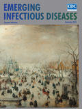
An Icy Vista from a Golden Age [PDF - 1.73 MB - 2 pages]
| EID | Breedlove B. An Icy Vista from a Golden Age. Emerg Infect Dis. 2018;24(12):2389-2390. https://doi.org/10.3201/eid2412.ac2412 |
|---|---|
| AMA | Breedlove B. An Icy Vista from a Golden Age. Emerging Infectious Diseases. 2018;24(12):2389-2390. doi:10.3201/eid2412.ac2412. |
| APA | Breedlove, B. (2018). An Icy Vista from a Golden Age. Emerging Infectious Diseases, 24(12), 2389-2390. https://doi.org/10.3201/eid2412.ac2412. |


