Synopses
Genotyping Approach for Potential Common Source of Enterocytozoon bieneusi Infection in Hematology Unit [PDF - 1.20 MB - 7 pages]
Microsporidiosis is a fungal infection that generally causes digestive disorders, especially in immunocompromised hosts. Over a 4-day period in January 2018, 3 patients with hematologic malignancies who were admitted to the hematology unit of a hospital in France received diagnoses of Enterocytozoon bieneusi microsporidiosis. This unusually high incidence was investigated by sequence analysis at the internal transcribed spacer rDNA locus and then by 3 microsatellites and 1 minisatellite for multilocus genotyping. The 3 isolates had many sequence similarities and belonged to a new genotype closely related to genotype C. In addition, multilocus genotyping showed high genetic distances with all the other strains collected from epidemiologically unrelated persons; none of these strains belonged to the new genotype. These data confirm the epidemiologic link among the 3 patients and support a common source of infection.
| EID | Desoubeaux G, Nourrisson C, Moniot M, De Kyvon M, Bonnin V, De La Bretonniére M, et al. Genotyping Approach for Potential Common Source of Enterocytozoon bieneusi Infection in Hematology Unit. Emerg Infect Dis. 2019;25(9):1625-1631. https://doi.org/10.3201/eid2509.190311 |
|---|---|
| AMA | Desoubeaux G, Nourrisson C, Moniot M, et al. Genotyping Approach for Potential Common Source of Enterocytozoon bieneusi Infection in Hematology Unit. Emerging Infectious Diseases. 2019;25(9):1625-1631. doi:10.3201/eid2509.190311. |
| APA | Desoubeaux, G., Nourrisson, C., Moniot, M., De Kyvon, M., Bonnin, V., De La Bretonniére, M....Poirier, P. (2019). Genotyping Approach for Potential Common Source of Enterocytozoon bieneusi Infection in Hematology Unit. Emerging Infectious Diseases, 25(9), 1625-1631. https://doi.org/10.3201/eid2509.190311. |
Epidemiology of Carbapenemase-Producing Klebsiella pneumoniae in a Hospital, Portugal [PDF - 913 KB - 6 pages]
We aimed to provide updated epidemiologic data on carbapenem-resistant Klebsiella pneumoniae in Portugal by characterizing all isolates (N = 46) recovered during 2013–2018 in a 123-bed hospital in Lisbon. We identified blaKPC-3 (n = 36), blaOXA-181 (n = 9), and blaGES-5 (n = 8) carbapenemase genes and observed co-occurrence of blaKPC-3 and blaGES-5 in 7 isolates. A single GES-5–producing isolate co-produced the extended-spectrum β-lactamase BEL-1; both corresponding genes were co-located on the same ColE1-like plasmid. The blaOXA-181 gene was always located on an IncX3 plasmid, whereas blaKPC-3 was carried on IncN, IncFII, IncFIB, and IncFIIA plasmid types. The 46 isolates were distributed into 13 pulsotypes and 9 sequence types. All isolates remained susceptible to ceftazidime/avibactam, but some exhibited reduced antimicrobial susceptibility (MIC = 3 mg/L).
| EID | Aires-de-Sousa M, Ortiz de la Rosa J, Gonçalves M, Pereira A, Nordmann P, Poirel L. Epidemiology of Carbapenemase-Producing Klebsiella pneumoniae in a Hospital, Portugal. Emerg Infect Dis. 2019;25(9):1632-1638. https://doi.org/10.3201/eid2509.190656 |
|---|---|
| AMA | Aires-de-Sousa M, Ortiz de la Rosa J, Gonçalves M, et al. Epidemiology of Carbapenemase-Producing Klebsiella pneumoniae in a Hospital, Portugal. Emerging Infectious Diseases. 2019;25(9):1632-1638. doi:10.3201/eid2509.190656. |
| APA | Aires-de-Sousa, M., Ortiz de la Rosa, J., Gonçalves, M., Pereira, A., Nordmann, P., & Poirel, L. (2019). Epidemiology of Carbapenemase-Producing Klebsiella pneumoniae in a Hospital, Portugal. Emerging Infectious Diseases, 25(9), 1632-1638. https://doi.org/10.3201/eid2509.190656. |
To evaluate a classification system to support clinical decisions for treatment of contaminated deep wounds at risk for an invasive fungal infection (IFI), we studied 246 US service members (413 wounds) injured in Afghanistan (2009–2014) who had laboratory evidence of fungal infection. A total of 143 wounds with persistent necrosis and laboratory evidence were classified as IFI; 120 wounds not meeting IFI criteria were classified as high suspicion (patients had localized infection signs/symptoms and had received antifungal medication for >10 days), and 150 were classified as low suspicion (failed to meet these criteria). IFI patients received more blood than other patients and had more severe injuries than patients in the low-suspicion group. Fungi of the order Mucorales were more frequently isolated from IFI (39%) and high-suspicion (21%) wounds than from low-suspicion (9%) wounds. Wounds that did not require immediate antifungal therapy lacked necrosis and localized signs/symptoms of infection and contained fungi from orders other than Mucorales.
| EID | Ganesan A, Shaikh F, Bradley W, Blyth DM, Bennett D, Petfield JL, et al. Classification of Trauma-Associated Invasive Fungal Infections to Support Wound Treatment Decisions. Emerg Infect Dis. 2019;25(9):1639-1647. https://doi.org/10.3201/eid2509.190168 |
|---|---|
| AMA | Ganesan A, Shaikh F, Bradley W, et al. Classification of Trauma-Associated Invasive Fungal Infections to Support Wound Treatment Decisions. Emerging Infectious Diseases. 2019;25(9):1639-1647. doi:10.3201/eid2509.190168. |
| APA | Ganesan, A., Shaikh, F., Bradley, W., Blyth, D. M., Bennett, D., Petfield, J. L....Tribble, D. R. (2019). Classification of Trauma-Associated Invasive Fungal Infections to Support Wound Treatment Decisions. Emerging Infectious Diseases, 25(9), 1639-1647. https://doi.org/10.3201/eid2509.190168. |
Clinical Characteristics and Treatment Outcomes for Patients Infected with Mycobacterium haemophilum [PDF - 1.13 MB - 5 pages]
Mycobacterium haemophilum is a nontuberculous mycobacterium that can infect immunocompromised patients. Because of special conditions required for its culture, this bacterium is rarely reported and there are scarce data for long-term outcomes. We conducted a retrospective study at Siriraj Hospital, Bangkok, Thailand, during January 2012–September 2017. We studied 21 patients for which HIV infection was the most common concurrent condition. The most common organ involvement was skin and soft tissue (60%). Combination therapy with macrolides and fluoroquinolones resulted in a 60% cure rate for cutaneous infection; adding rifampin as a third drug for more severe cases resulted in modest (66%) cure rate. Efficacy of medical therapy in cutaneous, musculoskeletal, and ocular diseases was 80%, 50%, and 50%, respectively. All patients with central nervous system involvement showed treatment failures. Infections with M. haemophilum in HIV-infected patients were more likely to have central nervous system involvement and tended to have disseminated infections and less favorable outcomes.
| EID | Nookeu P, Angkasekwinai N, Foongladda S, Phoompoung P. Clinical Characteristics and Treatment Outcomes for Patients Infected with Mycobacterium haemophilum. Emerg Infect Dis. 2019;25(9):1648-1652. https://doi.org/10.3201/eid2509.190430 |
|---|---|
| AMA | Nookeu P, Angkasekwinai N, Foongladda S, et al. Clinical Characteristics and Treatment Outcomes for Patients Infected with Mycobacterium haemophilum. Emerging Infectious Diseases. 2019;25(9):1648-1652. doi:10.3201/eid2509.190430. |
| APA | Nookeu, P., Angkasekwinai, N., Foongladda, S., & Phoompoung, P. (2019). Clinical Characteristics and Treatment Outcomes for Patients Infected with Mycobacterium haemophilum. Emerging Infectious Diseases, 25(9), 1648-1652. https://doi.org/10.3201/eid2509.190430. |
Research
Theileria orientalis Ikeda Genotype in Cattle, Virginia, USA [PDF - 1.00 MB - 7 pages]
Theileria orientalis Ikeda genotype is a parasite that causes a disease in cattle that results in major economic issues in Asia, New Zealand, and Australia. The parasite is transmitted by Haemaphysalis longicornis ticks, which have recently been reported in numerous states throughout the eastern United States. Concurrently, cattle in Virginia showed clinical signs consistent with a hemoprotozoan infection. We used amplicons specific for the major piroplasm surface protein and small subunit rDNA of piroplasms to test blood samples from the cattle by PCR. Bidirectional Sanger sequencing showed sequences with 100% identity with T. orientalis Ikeda genotype 2 sequences. We detected the parasite in 3 unrelated herds and from various animals sampled at 2 time points. Although other benign T. orientalis genotypes are endemic to the United States, detection of T. orientalis Ikeda genotype might represent a risk for the cattle industry in Virginia.
| EID | Oakes VJ, Yabsley MJ, Schwartz D, LeRoith T, Bissett C, Broaddus C, et al. Theileria orientalis Ikeda Genotype in Cattle, Virginia, USA. Emerg Infect Dis. 2019;25(9):1653-1659. https://doi.org/10.3201/eid2509.190088 |
|---|---|
| AMA | Oakes VJ, Yabsley MJ, Schwartz D, et al. Theileria orientalis Ikeda Genotype in Cattle, Virginia, USA. Emerging Infectious Diseases. 2019;25(9):1653-1659. doi:10.3201/eid2509.190088. |
| APA | Oakes, V. J., Yabsley, M. J., Schwartz, D., LeRoith, T., Bissett, C., Broaddus, C....Lahmers, K. K. (2019). Theileria orientalis Ikeda Genotype in Cattle, Virginia, USA. Emerging Infectious Diseases, 25(9), 1653-1659. https://doi.org/10.3201/eid2509.190088. |
Genetic Characterization and Enhanced Surveillance of Ceftriaxone-Resistant Neisseria gonorrhoeae Strain, Alberta, Canada, 2018 [PDF - 626 KB - 8 pages]
In July 2018, a case of Neisseria gonorrhoeae associated with ceftriaxone treatment failure was identified in Alberta, Canada. We identified the isolate and nucleic acid amplification testing (NAAT) specimen as the ceftriaxone-resistant strain multilocus sequence type 1903/NG-MAST 3435/NG-STAR 233, originally identified in Japan (FC428), with the same penA 60.001 mosaic allele and genetic resistance determinants. Core single-nucleotide variant (SNV) analysis identified 13 SNVs between this isolate and FC428. Culture-independent surveillance by PCR for the A311V mutation in the penA allele and N. gonorrhoeae multiantigen sequence typing directly from NAAT transport media positive for N. gonorrhoeae by NAAT did not detect spread of the strain. We identified multiple sequence types not previously detected in Alberta by routine surveillance. This case demonstrates the benefit of using culture-independent methods to enhance detection, public health investigations, and surveillance to address this global threat.
| EID | Berenger BM, Demczuk W, Gratrix J, Pabbaraju K, Smyczek P, Martin I. Genetic Characterization and Enhanced Surveillance of Ceftriaxone-Resistant Neisseria gonorrhoeae Strain, Alberta, Canada, 2018. Emerg Infect Dis. 2019;25(9):1660-1667. https://doi.org/10.3201/eid2509.190407 |
|---|---|
| AMA | Berenger BM, Demczuk W, Gratrix J, et al. Genetic Characterization and Enhanced Surveillance of Ceftriaxone-Resistant Neisseria gonorrhoeae Strain, Alberta, Canada, 2018. Emerging Infectious Diseases. 2019;25(9):1660-1667. doi:10.3201/eid2509.190407. |
| APA | Berenger, B. M., Demczuk, W., Gratrix, J., Pabbaraju, K., Smyczek, P., & Martin, I. (2019). Genetic Characterization and Enhanced Surveillance of Ceftriaxone-Resistant Neisseria gonorrhoeae Strain, Alberta, Canada, 2018. Emerging Infectious Diseases, 25(9), 1660-1667. https://doi.org/10.3201/eid2509.190407. |
Clonality of Fluconazole-Nonsusceptible Candida tropicalis in Bloodstream Infections, Taiwan, 2011–2017 [PDF - 985 KB - 8 pages]
Candida tropicalis is the leading cause of non–C. albicans candidemia in tropical Asia and Latin America. We evaluated isolates from 344 patients with an initial episode of C. tropicalis candidemia. We found that 58 (16.9%) patients were infected by fluconazole-nonsusceptible (FNS) C. tropicalis with cross resistance to itraconazole, voriconazole, and posaconazole; 55.2% (32/58) of patients were azole-naive. Multilocus sequence typing analysis revealed FNS isolates were genetically closely related, but we did not see time- or place-clustering. Among the diploid sequence types (DSTs), we noted DST225, which has been reported from fruit in Taiwan and hospitals in Beijing, China, as well as DST376 and DST505–7, which also were reported from hospitals in Shanghai, China. Our findings suggest cross-boundary expansion of FNS C. tropicalis and highlight the importance of active surveillance of clinical isolates to detect dissemination of this pathogen and explore potential sources in the community.
| EID | Chen P, Chuang Y, Wu U, Sun H, Wang J, Sheng W, et al. Clonality of Fluconazole-Nonsusceptible Candida tropicalis in Bloodstream Infections, Taiwan, 2011–2017. Emerg Infect Dis. 2019;25(9):1668-1675. https://doi.org/10.3201/eid2509.190520 |
|---|---|
| AMA | Chen P, Chuang Y, Wu U, et al. Clonality of Fluconazole-Nonsusceptible Candida tropicalis in Bloodstream Infections, Taiwan, 2011–2017. Emerging Infectious Diseases. 2019;25(9):1668-1675. doi:10.3201/eid2509.190520. |
| APA | Chen, P., Chuang, Y., Wu, U., Sun, H., Wang, J., Sheng, W....Chang, S. (2019). Clonality of Fluconazole-Nonsusceptible Candida tropicalis in Bloodstream Infections, Taiwan, 2011–2017. Emerging Infectious Diseases, 25(9), 1668-1675. https://doi.org/10.3201/eid2509.190520. |
Association of Enterovirus D68 with Acute Flaccid Myelitis, Philadelphia, Pennsylvania, USA, 2009–2018 [PDF - 1.32 MB - 7 pages]
Acute flaccid myelitis (AFM) is a polio-like disease that results in paralysis in previously healthy persons. Although the definitive cause of AFM remains unconfirmed, enterovirus D68 (EV-D68) is suspected based on 2014 data demonstrating an increase in AFM cases concomitant with an EV-D68 outbreak. We examined the prevalence in children and the molecular evolution of EV-D68 for 2009–2018 in Philadelphia, Pennsylvania, USA. We detected widespread EV-D68 circulation in 2009, rare detections in 2010 and 2011, and then biennial circulation, only in even years, during 2012–2018. Prevalence of EV-D68 significantly correlated with AFM cases during this period. Finally, whole-genome sequencing revealed early detection of the B1 clade in 2009 and continued evolution of the B3 clade from 2016 to 2018. These data reinforce the need to improve surveillance programs for nonpolio enterovirus to identify possible AFM triggers and predict disease prevalence to better prepare for future outbreaks.
| EID | Uprety P, Curtis D, Elkan M, Fink J, Rajagopalan R, Zhao C, et al. Association of Enterovirus D68 with Acute Flaccid Myelitis, Philadelphia, Pennsylvania, USA, 2009–2018. Emerg Infect Dis. 2019;25(9):1676-1682. https://doi.org/10.3201/eid2509.190468 |
|---|---|
| AMA | Uprety P, Curtis D, Elkan M, et al. Association of Enterovirus D68 with Acute Flaccid Myelitis, Philadelphia, Pennsylvania, USA, 2009–2018. Emerging Infectious Diseases. 2019;25(9):1676-1682. doi:10.3201/eid2509.190468. |
| APA | Uprety, P., Curtis, D., Elkan, M., Fink, J., Rajagopalan, R., Zhao, C....Graf, E. H. (2019). Association of Enterovirus D68 with Acute Flaccid Myelitis, Philadelphia, Pennsylvania, USA, 2009–2018. Emerging Infectious Diseases, 25(9), 1676-1682. https://doi.org/10.3201/eid2509.190468. |
To assess whether risk for Clostridioides difficile infection (CDI) is higher among older adults with cancer, we conducted a retrospective cohort study with a nested case–control analysis using population-based Surveillance, Epidemiology, and End Results–Medicare linked data for 2011. Among 93,566 Medicare beneficiaries, incident CDI and odds for acquiring CDI were higher among patients with than without cancer. Specifically, risk was significantly higher for those who had liquid tumors and higher for those who had recently diagnosed solid tumors and distant metastasis. These findings were independent of prior healthcare-associated exposure. This population-based assessment can be used to identify targets for prevention of CDI.
| EID | Kamboj M, Gennarelli RL, Brite J, Sepkowitz K, Lipitz-Snyderman A. Risk for Clostridioides difficile Infection among Older Adults with Cancer. Emerg Infect Dis. 2019;25(9):1683-1689. https://doi.org/10.3201/eid2509.181142 |
|---|---|
| AMA | Kamboj M, Gennarelli RL, Brite J, et al. Risk for Clostridioides difficile Infection among Older Adults with Cancer. Emerging Infectious Diseases. 2019;25(9):1683-1689. doi:10.3201/eid2509.181142. |
| APA | Kamboj, M., Gennarelli, R. L., Brite, J., Sepkowitz, K., & Lipitz-Snyderman, A. (2019). Risk for Clostridioides difficile Infection among Older Adults with Cancer. Emerging Infectious Diseases, 25(9), 1683-1689. https://doi.org/10.3201/eid2509.181142. |
Whole-Genome Sequencing of Salmonella Mississippi and Typhimurium Definitive Type 160, Australia and New Zealand [PDF - 2.68 MB - 8 pages]
We used phylogenomic and risk factor data on isolates of Salmonella enterica serovars Mississippi and Typhimurium definitive type 160 (DT160) collected from human, animal, and environmental sources to elucidate their epidemiology and disease reservoirs in Australia and New Zealand. Sequence data suggested wild birds as a likely reservoir for DT160; animal and environmental sources varied more for Salmonella Mississippi than for Salmonella Typhimurium. Australia and New Zealand isolates sat in distinct clades for both serovars; the median single-nucleotide polymorphism distance for DT160 was 29 (range 8–66) and for Salmonella Mississippi, 619 (range 565–737). Phylogenomic data identified plausible sources of human infection from wildlife and environmental reservoirs and provided evidence supporting New Zealand–acquired DT160 in a group of travelers returning to Australia. Wider use of real-time whole-genome sequencing in new locations and for other serovars may identify sources and routes of transmission, thereby aiding prevention and control.
| EID | Ford L, Ingle D, Glass K, Veitch M, Williamson DA, Harlock M, et al. Whole-Genome Sequencing of Salmonella Mississippi and Typhimurium Definitive Type 160, Australia and New Zealand. Emerg Infect Dis. 2019;25(9):1690-1697. https://doi.org/10.3201/eid2509.181811 |
|---|---|
| AMA | Ford L, Ingle D, Glass K, et al. Whole-Genome Sequencing of Salmonella Mississippi and Typhimurium Definitive Type 160, Australia and New Zealand. Emerging Infectious Diseases. 2019;25(9):1690-1697. doi:10.3201/eid2509.181811. |
| APA | Ford, L., Ingle, D., Glass, K., Veitch, M., Williamson, D. A., Harlock, M....Kirk, M. D. (2019). Whole-Genome Sequencing of Salmonella Mississippi and Typhimurium Definitive Type 160, Australia and New Zealand. Emerging Infectious Diseases, 25(9), 1690-1697. https://doi.org/10.3201/eid2509.181811. |
Epidemiologic Shift in Candidemia Driven by Candida auris, South Africa, 2016–2017 [PDF - 2.00 MB - 10 pages]
Candida auris is an invasive healthcare-associated fungal pathogen. Cases of candidemia, defined as illness in patients with Candida cultured from blood, were detected through national laboratory-based surveillance in South Africa during 2016–2017. We identified viable isolates by using mass spectrometry and sequencing. Among 6,669 cases (5,876 with species identification) from 269 hospitals, 794 (14%) were caused by C. auris. The incidence risk for all candidemia at 133 hospitals was 83.8 (95% CI 81.2–86.4) cases/100,000 admissions. Prior systemic antifungal drug therapy was associated with a 40% increased adjusted odds of C. auris fungemia compared with bloodstream infection caused by other Candida species (adjusted odds ratio 1.4 [95% CI 0.8–2.3]). The crude in-hospital case-fatality ratio did not differ between Candida species and was 45% for C. auris candidemia, compared with 43% for non–C. auris candidemia. C. auris has caused a major epidemiologic shift in candidemia in South Africa.
| EID | van Schalkwyk E, Mpembe RS, Thomas J, Shuping L, Ismail H, Lowman W, et al. Epidemiologic Shift in Candidemia Driven by Candida auris, South Africa, 2016–2017. Emerg Infect Dis. 2019;25(9):1698-1707. https://doi.org/10.3201/eid2509.190040 |
|---|---|
| AMA | van Schalkwyk E, Mpembe RS, Thomas J, et al. Epidemiologic Shift in Candidemia Driven by Candida auris, South Africa, 2016–2017. Emerging Infectious Diseases. 2019;25(9):1698-1707. doi:10.3201/eid2509.190040. |
| APA | van Schalkwyk, E., Mpembe, R. S., Thomas, J., Shuping, L., Ismail, H., Lowman, W....Govender, N. P. (2019). Epidemiologic Shift in Candidemia Driven by Candida auris, South Africa, 2016–2017. Emerging Infectious Diseases, 25(9), 1698-1707. https://doi.org/10.3201/eid2509.190040. |
Effect of Pneumococcal Conjugate Vaccines on Pneumococcal Meningitis, England and Wales, July 1, 2000–June 30, 2016 [PDF - 2.43 MB - 11 pages]
We describe the effects of the 7-valent (PCV7) and 13-valent (PCV13) pneumococcal conjugate vaccines on pneumococcal meningitis in England and Wales during July 1, 2000–June 30, 2016. Overall, 84,473 laboratory-confirmed invasive pneumococcal disease cases, including 4,160 (4.9%) cases with meningitis, occurred. PCV7 implementation in 2006 did not lower overall pneumococcal meningitis incidence because of replacement with non–PCV7-type meningitis incidence. Replacement with PCV13 in 2010, however, led to a 48% reduction in pneumococcal meningitis incidence by 2015–16. The overall case-fatality rate was 17.5%: 10.7% among patients <5 years of age, 17.3% among patients 5–64 years of age, and 31.9% among patients >65 years of age. Serotype 8 was associated with increased odds of death (adjusted odds ratio 2.9, 95% CI 1.8–4.7). In England and Wales, an effect on pneumococcal meningitis was observed only after PCV13 implementation. Further studies are needed to assess pneumococcal meningitis caused by the replacing serotypes.
| EID | Oligbu G, Collins S, Djennad A, Sheppard CL, Fry NK, Andrews NJ, et al. Effect of Pneumococcal Conjugate Vaccines on Pneumococcal Meningitis, England and Wales, July 1, 2000–June 30, 2016. Emerg Infect Dis. 2019;25(9):1708-1718. https://doi.org/10.3201/eid2509.180747 |
|---|---|
| AMA | Oligbu G, Collins S, Djennad A, et al. Effect of Pneumococcal Conjugate Vaccines on Pneumococcal Meningitis, England and Wales, July 1, 2000–June 30, 2016. Emerging Infectious Diseases. 2019;25(9):1708-1718. doi:10.3201/eid2509.180747. |
| APA | Oligbu, G., Collins, S., Djennad, A., Sheppard, C. L., Fry, N. K., Andrews, N. J....Ladhani, S. N. (2019). Effect of Pneumococcal Conjugate Vaccines on Pneumococcal Meningitis, England and Wales, July 1, 2000–June 30, 2016. Emerging Infectious Diseases, 25(9), 1708-1718. https://doi.org/10.3201/eid2509.180747. |
Dispatches
Rickettsia japonica Infections in Humans, Xinyang, China, 2014–2017 [PDF - 2.50 MB - 4 pages]
During 2014–2017, we screened for Rickettsia japonica infection in Xinyang, China, and identified 20 cases. The major clinical manifestations of monoinfection were fever, asthenia, myalgia, rash, and anorexia; laboratory findings included thrombocytopenia and elevated hepatic aminotransferase concentrations. Physicians in China should consider R. japonica infection in at-risk patients.
| EID | Li H, Zhang P, Du J, Yang Z, Cui N, Xing B, et al. Rickettsia japonica Infections in Humans, Xinyang, China, 2014–2017. Emerg Infect Dis. 2019;25(9):1719-1722. https://doi.org/10.3201/eid2509.171421 |
|---|---|
| AMA | Li H, Zhang P, Du J, et al. Rickettsia japonica Infections in Humans, Xinyang, China, 2014–2017. Emerging Infectious Diseases. 2019;25(9):1719-1722. doi:10.3201/eid2509.171421. |
| APA | Li, H., Zhang, P., Du, J., Yang, Z., Cui, N., Xing, B....Liu, W. (2019). Rickettsia japonica Infections in Humans, Xinyang, China, 2014–2017. Emerging Infectious Diseases, 25(9), 1719-1722. https://doi.org/10.3201/eid2509.171421. |
Climate Classification System–Based Determination of Temperate Climate Detection of Cryptococcus gattii sensu lato [PDF - 1.28 MB - 4 pages]
We compared 2 climate classification systems describing georeferenced environmental Cryptococcus gattii sensu lato isolations occurring during 1989–2016. Each system suggests the fungus was isolated in temperate climates before the 1999 outbreak on Vancouver Island, British Columbia, Canada. However, the Köppen-Geiger system is more precise and should be used to define climates where pathogens are detected.
| EID | Acheson ES, Galanis E, Bartlett K, Klinkenberg B. Climate Classification System–Based Determination of Temperate Climate Detection of Cryptococcus gattii sensu lato. Emerg Infect Dis. 2019;25(9):1723-1726. https://doi.org/10.3201/eid2509.181884 |
|---|---|
| AMA | Acheson ES, Galanis E, Bartlett K, et al. Climate Classification System–Based Determination of Temperate Climate Detection of Cryptococcus gattii sensu lato. Emerging Infectious Diseases. 2019;25(9):1723-1726. doi:10.3201/eid2509.181884. |
| APA | Acheson, E. S., Galanis, E., Bartlett, K., & Klinkenberg, B. (2019). Climate Classification System–Based Determination of Temperate Climate Detection of Cryptococcus gattii sensu lato. Emerging Infectious Diseases, 25(9), 1723-1726. https://doi.org/10.3201/eid2509.181884. |
Cluster of Nasal Rhinosporidiosis, Eastern Province, Rwanda [PDF - 929 KB - 3 pages]
We report 4 recent cases of nasal rhinosporidiosis in Rwanda. All patients were boys or young men living in the same district (Gatsibo District, Eastern Province), suggesting a reservoir in the area. The recent reemergence of rhinosporidiosis in Rwanda might reflect increased availability of diagnostic services rather than emerging disease.
| EID | Izimukwiye AI, Mbarushimana D, Ndayisaba MC, Bigirimana V, Rugwizangoga B, Laga AC. Cluster of Nasal Rhinosporidiosis, Eastern Province, Rwanda. Emerg Infect Dis. 2019;25(9):1727-1729. https://doi.org/10.3201/eid2509.190021 |
|---|---|
| AMA | Izimukwiye AI, Mbarushimana D, Ndayisaba MC, et al. Cluster of Nasal Rhinosporidiosis, Eastern Province, Rwanda. Emerging Infectious Diseases. 2019;25(9):1727-1729. doi:10.3201/eid2509.190021. |
| APA | Izimukwiye, A. I., Mbarushimana, D., Ndayisaba, M. C., Bigirimana, V., Rugwizangoga, B., & Laga, A. C. (2019). Cluster of Nasal Rhinosporidiosis, Eastern Province, Rwanda. Emerging Infectious Diseases, 25(9), 1727-1729. https://doi.org/10.3201/eid2509.190021. |
Use of Human Intestinal Enteroids to Detect Human Norovirus Infectivity [PDF - 1.04 MB - 6 pages]
Tools to detect human norovirus infectivity have been lacking. Using human intestinal enteroid cultures inoculated with GII.Pe-GII.4 Sydney–infected fecal samples, we determined that a real-time reverse transcription PCR cycle threshold cutoff of 30 may indicate infectious norovirus. This finding could be used to help guide infection control.
| EID | Chan M, Cheung S, Mohammad KN, Chan J, Estes MK, Chan P. Use of Human Intestinal Enteroids to Detect Human Norovirus Infectivity. Emerg Infect Dis. 2019;25(9):1730-1735. https://doi.org/10.3201/eid2509.190205 |
|---|---|
| AMA | Chan M, Cheung S, Mohammad KN, et al. Use of Human Intestinal Enteroids to Detect Human Norovirus Infectivity. Emerging Infectious Diseases. 2019;25(9):1730-1735. doi:10.3201/eid2509.190205. |
| APA | Chan, M., Cheung, S., Mohammad, K. N., Chan, J., Estes, M. K., & Chan, P. (2019). Use of Human Intestinal Enteroids to Detect Human Norovirus Infectivity. Emerging Infectious Diseases, 25(9), 1730-1735. https://doi.org/10.3201/eid2509.190205. |
Vaccine Effectiveness against DS-1–Like Rotavirus Strains in Infants with Acute Gastroenteritis, Malawi, 2013–2015 [PDF - 881 KB - 4 pages]
Atypical DS-1–like G1P[8] rotaviruses emerged in 2013 in Malawi after rotavirus vaccine introduction. Vaccine effectiveness among infants hospitalized with acute DS-1–like G1P[8] rotavirus gastroenteritis was 85.6% (95% CI 34.4%–96.8%). These findings suggest that vaccine provides protection against these strains despite their emergence coinciding with vaccine introduction.
| EID | Jere KC, Bar-Zeev N, Chande A, Bennett A, Pollock L, Sanchez-Lopez PF, et al. Vaccine Effectiveness against DS-1–Like Rotavirus Strains in Infants with Acute Gastroenteritis, Malawi, 2013–2015. Emerg Infect Dis. 2019;25(9):1734-1737. https://doi.org/10.3201/eid2509.190258 |
|---|---|
| AMA | Jere KC, Bar-Zeev N, Chande A, et al. Vaccine Effectiveness against DS-1–Like Rotavirus Strains in Infants with Acute Gastroenteritis, Malawi, 2013–2015. Emerging Infectious Diseases. 2019;25(9):1734-1737. doi:10.3201/eid2509.190258. |
| APA | Jere, K. C., Bar-Zeev, N., Chande, A., Bennett, A., Pollock, L., Sanchez-Lopez, P. F....Cunliffe, N. A. (2019). Vaccine Effectiveness against DS-1–Like Rotavirus Strains in Infants with Acute Gastroenteritis, Malawi, 2013–2015. Emerging Infectious Diseases, 25(9), 1734-1737. https://doi.org/10.3201/eid2509.190258. |
Rodent Host Abundance and Climate Variability as Predictors of Tickborne Disease Risk 1 Year in Advance [PDF - 1.86 MB - 4 pages]
Using long-term data on incidences of Lyme disease and tickborne encephalitis, we showed that the dynamics of both diseases in central Europe are predictable from rodent host densities and climate indices. Our approach offers a simple and effective tool to predict a tickborne disease risk 1 year in advance.
| EID | Tkadlec E, Václavík T, Široký P. Rodent Host Abundance and Climate Variability as Predictors of Tickborne Disease Risk 1 Year in Advance. Emerg Infect Dis. 2019;25(9):1738-1741. https://doi.org/10.3201/eid2509.190684 |
|---|---|
| AMA | Tkadlec E, Václavík T, Široký P. Rodent Host Abundance and Climate Variability as Predictors of Tickborne Disease Risk 1 Year in Advance. Emerging Infectious Diseases. 2019;25(9):1738-1741. doi:10.3201/eid2509.190684. |
| APA | Tkadlec, E., Václavík, T., & Široký, P. (2019). Rodent Host Abundance and Climate Variability as Predictors of Tickborne Disease Risk 1 Year in Advance. Emerging Infectious Diseases, 25(9), 1738-1741. https://doi.org/10.3201/eid2509.190684. |
Delays in Coccidioidomycosis Diagnosis and Relationship to Healthcare Utilization, Phoenix, Arizona, USA [PDF - 367 KB - 3 pages]
We developed an electronic records methodology to programmatically estimate the date of first appearance of coccidioidomycosis symptoms in patients. We compared the diagnostic delay with overall healthcare utilization charges. Many patients (46%) had delays in diagnosis of >1 month. Billed healthcare charges before diagnosis increased with length of delay.
| EID | Ginn R, Mohty R, Bollmann K, Goodsell J, Mendez G, Bradley B, et al. Delays in Coccidioidomycosis Diagnosis and Relationship to Healthcare Utilization, Phoenix, Arizona, USA. Emerg Infect Dis. 2019;25(9):1742-1744. https://doi.org/10.3201/eid2509.190019 |
|---|---|
| AMA | Ginn R, Mohty R, Bollmann K, et al. Delays in Coccidioidomycosis Diagnosis and Relationship to Healthcare Utilization, Phoenix, Arizona, USA. Emerging Infectious Diseases. 2019;25(9):1742-1744. doi:10.3201/eid2509.190019. |
| APA | Ginn, R., Mohty, R., Bollmann, K., Goodsell, J., Mendez, G., Bradley, B....Galgiani, J. N. (2019). Delays in Coccidioidomycosis Diagnosis and Relationship to Healthcare Utilization, Phoenix, Arizona, USA. Emerging Infectious Diseases, 25(9), 1742-1744. https://doi.org/10.3201/eid2509.190019. |
Delays in Coccidioidomycosis Diagnosis and Associated Healthcare Utilization, Tucson, Arizona, USA [PDF - 436 KB - 3 pages]
Tucson, Arizona, USA, is a highly coccidioidomycosis-endemic area. We conducted a retrospective review of 815 patients in Tucson over 2.7 years. Of 276 patients with coccidioidomycosis, 246 had a delay in diagnosis; median delay was 23 days. Diagnosis delay was associated with coccidioidomycosis-related costs totaling $589,053 and included extensive antibacterial drug use.
| EID | Donovan FM, Wightman P, Zong Y, Gabe L, Majeed A, Ynosencio T, et al. Delays in Coccidioidomycosis Diagnosis and Associated Healthcare Utilization, Tucson, Arizona, USA. Emerg Infect Dis. 2019;25(9):1745-1747. https://doi.org/10.3201/eid2509.190023 |
|---|---|
| AMA | Donovan FM, Wightman P, Zong Y, et al. Delays in Coccidioidomycosis Diagnosis and Associated Healthcare Utilization, Tucson, Arizona, USA. Emerging Infectious Diseases. 2019;25(9):1745-1747. doi:10.3201/eid2509.190023. |
| APA | Donovan, F. M., Wightman, P., Zong, Y., Gabe, L., Majeed, A., Ynosencio, T....Galgiani, J. N. (2019). Delays in Coccidioidomycosis Diagnosis and Associated Healthcare Utilization, Tucson, Arizona, USA. Emerging Infectious Diseases, 25(9), 1745-1747. https://doi.org/10.3201/eid2509.190023. |
Research Letters
Soft Tissue Infection with Diaporthe phaseolorum in Heart Transplant Recipient with End-Stage Renal Failure [PDF - 744 KB - 2 pages]
Diaporthe phaseolorum is a fungal plant parasite that has rarely been described as causing invasive human disease. We report a case of human soft tissue infection with Diaporthe phaseolorum in a heart transplant patient with end-stage renal failure in New Zealand.
| EID | Howard JC, Chen K, Werno A, Metcalf S. Soft Tissue Infection with Diaporthe phaseolorum in Heart Transplant Recipient with End-Stage Renal Failure. Emerg Infect Dis. 2019;25(9):1748-1749. https://doi.org/10.3201/eid2509.190768 |
|---|---|
| AMA | Howard JC, Chen K, Werno A, et al. Soft Tissue Infection with Diaporthe phaseolorum in Heart Transplant Recipient with End-Stage Renal Failure. Emerging Infectious Diseases. 2019;25(9):1748-1749. doi:10.3201/eid2509.190768. |
| APA | Howard, J. C., Chen, K., Werno, A., & Metcalf, S. (2019). Soft Tissue Infection with Diaporthe phaseolorum in Heart Transplant Recipient with End-Stage Renal Failure. Emerging Infectious Diseases, 25(9), 1748-1749. https://doi.org/10.3201/eid2509.190768. |
Disseminated Emergomycosis in a Person with HIV Infection, Uganda [PDF - 993 KB - 2 pages]
We describe emergomycosis in a patient in Uganda with HIV infection. We tested a formalin-fixed, paraffin-embedded skin biopsy to identify Emergomyces pasteurianus or a closely related pathogen by sequencing broad-range fungal PCR amplicons. Results suggest that emergomycosis is more widespread and genetically diverse than previously documented. PCR on tissue blocks may help clarify emergomycosis epidemiology.
| EID | Rooms I, Mugisha P, Gambichler T, Hadaschik E, Esser S, Rath P, et al. Disseminated Emergomycosis in a Person with HIV Infection, Uganda. Emerg Infect Dis. 2019;25(9):1750-1751. https://doi.org/10.3201/eid2509.181234 |
|---|---|
| AMA | Rooms I, Mugisha P, Gambichler T, et al. Disseminated Emergomycosis in a Person with HIV Infection, Uganda. Emerging Infectious Diseases. 2019;25(9):1750-1751. doi:10.3201/eid2509.181234. |
| APA | Rooms, I., Mugisha, P., Gambichler, T., Hadaschik, E., Esser, S., Rath, P....Rickerts, V. (2019). Disseminated Emergomycosis in a Person with HIV Infection, Uganda. Emerging Infectious Diseases, 25(9), 1750-1751. https://doi.org/10.3201/eid2509.181234. |
Bourbon Virus in Wild and Domestic Animals, Missouri, USA, 2012–2013 [PDF - 353 KB - 2 pages]
Since its recent discovery, Bourbon virus has been isolated from a human and ticks. To assess exposure of potential vertebrate reservoirs, we assayed banked serum and plasma samples from wildlife and domestic animals in Missouri, USA, for Bourbon virus–neutralizing antibodies. We detected high seroprevalence in raccoons (50%) and white-tailed deer (86%).
| EID | Jackson KC, Gidlewski T, Root J, Bosco-Lauth AM, Lash R, Harmon JR, et al. Bourbon Virus in Wild and Domestic Animals, Missouri, USA, 2012–2013. Emerg Infect Dis. 2019;25(9):1752-1753. https://doi.org/10.3201/eid2509.181902 |
|---|---|
| AMA | Jackson KC, Gidlewski T, Root J, et al. Bourbon Virus in Wild and Domestic Animals, Missouri, USA, 2012–2013. Emerging Infectious Diseases. 2019;25(9):1752-1753. doi:10.3201/eid2509.181902. |
| APA | Jackson, K. C., Gidlewski, T., Root, J., Bosco-Lauth, A. M., Lash, R., Harmon, J. R....Komar, N. (2019). Bourbon Virus in Wild and Domestic Animals, Missouri, USA, 2012–2013. Emerging Infectious Diseases, 25(9), 1752-1753. https://doi.org/10.3201/eid2509.181902. |
Fatal Case of Lassa Fever, Bangolo District, Côte d’Ivoire, 2015 [PDF - 594 KB - 4 pages]
Lassa fever has not been reported in Côte d’Ivoire. We performed a retrospective analysis of human serum samples collected in Côte d’Ivoire in the dry seasons (January–April) during 2015–2018. We identified a fatal human case of Lassa fever in the Bangolo District of western Côte d’Ivoire during 2015.
| EID | Mateo M, Picard C, Sylla Y, Kamo E, Odegue D, Journeaux A, et al. Fatal Case of Lassa Fever, Bangolo District, Côte d’Ivoire, 2015. Emerg Infect Dis. 2019;25(9):1753-1756. https://doi.org/10.3201/eid2509.190239 |
|---|---|
| AMA | Mateo M, Picard C, Sylla Y, et al. Fatal Case of Lassa Fever, Bangolo District, Côte d’Ivoire, 2015. Emerging Infectious Diseases. 2019;25(9):1753-1756. doi:10.3201/eid2509.190239. |
| APA | Mateo, M., Picard, C., Sylla, Y., Kamo, E., Odegue, D., Journeaux, A....Dosso, M. (2019). Fatal Case of Lassa Fever, Bangolo District, Côte d’Ivoire, 2015. Emerging Infectious Diseases, 25(9), 1753-1756. https://doi.org/10.3201/eid2509.190239. |
Household Transmission of Human Adenovirus Type 55 in Case of Fatal Acute Respiratory Disease [PDF - 348 KB - 3 pages]
We identified a case of fatal acute respiratory disease from household transmission of human adenovirus type 55 (HAdV-55) in Anhui Province, China. Computed tomography showed severe pneumonia. Comparative genomic analysis of HAdV-55 indicated the virus possibly originated in Shanxi Province, China. More attention should be paid to highly contagious HAdV-55.
| EID | Jing S, Zhang J, Cao M, Liu M, Yan Y, Zhao S, et al. Household Transmission of Human Adenovirus Type 55 in Case of Fatal Acute Respiratory Disease. Emerg Infect Dis. 2019;25(9):1756-1758. https://doi.org/10.3201/eid2509.181937 |
|---|---|
| AMA | Jing S, Zhang J, Cao M, et al. Household Transmission of Human Adenovirus Type 55 in Case of Fatal Acute Respiratory Disease. Emerging Infectious Diseases. 2019;25(9):1756-1758. doi:10.3201/eid2509.181937. |
| APA | Jing, S., Zhang, J., Cao, M., Liu, M., Yan, Y., Zhao, S....Zhang, Q. (2019). Household Transmission of Human Adenovirus Type 55 in Case of Fatal Acute Respiratory Disease. Emerging Infectious Diseases, 25(9), 1756-1758. https://doi.org/10.3201/eid2509.181937. |
Worldwide Reduction in MERS Cases and Deaths since 2016 [PDF - 332 KB - 3 pages]
Since 2012, Middle East respiratory syndrome (MERS) coronavirus has infected 2,442 persons worldwide. Case-based data analysis suggests that since 2016, as many as 1,465 cases and 293–520 deaths might have been averted. Efforts to reduce the global MERS threat are working, but countries must maintain vigilance to prevent further infections.
| EID | Donnelly CA, Malik MR, Elkholy A, Cauchemez S, Van Kerkhove MD. Worldwide Reduction in MERS Cases and Deaths since 2016. Emerg Infect Dis. 2019;25(9):1758-1760. https://doi.org/10.3201/eid2509.190143 |
|---|---|
| AMA | Donnelly CA, Malik MR, Elkholy A, et al. Worldwide Reduction in MERS Cases and Deaths since 2016. Emerging Infectious Diseases. 2019;25(9):1758-1760. doi:10.3201/eid2509.190143. |
| APA | Donnelly, C. A., Malik, M. R., Elkholy, A., Cauchemez, S., & Van Kerkhove, M. D. (2019). Worldwide Reduction in MERS Cases and Deaths since 2016. Emerging Infectious Diseases, 25(9), 1758-1760. https://doi.org/10.3201/eid2509.190143. |
Limited Scope of Shorter Drug Regimen for MDR TB Caused by High Resistance to Fluoroquinolone [PDF - 428 KB - 3 pages]
Resistance to second-line tuberculosis drugs for patients with multidrug-resistant tuberculosis has emerged globally and is a potential risk factor for unfavorable outcomes of shorter duration drug regimens. We assessed the proportion of patients eligible for a shorter drug regimen in Uttar Pradesh, India, which had the highest rate of multidrug-resistant tuberculosis in India.
| EID | Singh PK, Jain A. Limited Scope of Shorter Drug Regimen for MDR TB Caused by High Resistance to Fluoroquinolone. Emerg Infect Dis. 2019;25(9):1760-1762. https://doi.org/10.3201/eid2509.190105 |
|---|---|
| AMA | Singh PK, Jain A. Limited Scope of Shorter Drug Regimen for MDR TB Caused by High Resistance to Fluoroquinolone. Emerging Infectious Diseases. 2019;25(9):1760-1762. doi:10.3201/eid2509.190105. |
| APA | Singh, P. K., & Jain, A. (2019). Limited Scope of Shorter Drug Regimen for MDR TB Caused by High Resistance to Fluoroquinolone. Emerging Infectious Diseases, 25(9), 1760-1762. https://doi.org/10.3201/eid2509.190105. |
Candida auris in Germany and Previous Exposure to Foreign Healthcare [PDF - 928 KB - 3 pages]
The emerging yeast Candida auris has disseminated worldwide. We report on 7 cases identified in Germany during 2015–2017. In 6 of these cases, C. auris was isolated from patients previously hospitalized abroad. Whole-genome sequencing and epidemiologic analyses revealed that all patients in Germany were infected with different strains.
| EID | Hamprecht A, Barber AE, Mellinghoff SC, Thelen P, Walther G, Yu Y, et al. Candida auris in Germany and Previous Exposure to Foreign Healthcare. Emerg Infect Dis. 2019;25(9):1763-1765. https://doi.org/10.3201/eid2509.190262 |
|---|---|
| AMA | Hamprecht A, Barber AE, Mellinghoff SC, et al. Candida auris in Germany and Previous Exposure to Foreign Healthcare. Emerging Infectious Diseases. 2019;25(9):1763-1765. doi:10.3201/eid2509.190262. |
| APA | Hamprecht, A., Barber, A. E., Mellinghoff, S. C., Thelen, P., Walther, G., Yu, Y....Kurzai, O. (2019). Candida auris in Germany and Previous Exposure to Foreign Healthcare. Emerging Infectious Diseases, 25(9), 1763-1765. https://doi.org/10.3201/eid2509.190262. |
Characterization of Clinical Isolates of Talaromyces marneffei and Related Species, California, USA [PDF - 594 KB - 4 pages]
Talaromyces marneffei and other Talaromyces species can cause opportunistic invasive fungal infections. We characterized clinical Talaromyces isolates from patients in California, USA, a non–Talaromyces-endemic area, by a multiphasic approach, including multigene phylogeny, matrix-assisted laser desorption/ionization time-of-flight mass spectrometry, and phenotypic methods. We identified 10 potentially pathogenic Talaromyces isolates, 2 T. marneffei.
| EID | Li L, Chen K, Dhungana N, Jang Y, Chaturvedi V, Desmond E. Characterization of Clinical Isolates of Talaromyces marneffei and Related Species, California, USA. Emerg Infect Dis. 2019;25(9):1765-1768. https://doi.org/10.3201/eid2509.190380 |
|---|---|
| AMA | Li L, Chen K, Dhungana N, et al. Characterization of Clinical Isolates of Talaromyces marneffei and Related Species, California, USA. Emerging Infectious Diseases. 2019;25(9):1765-1768. doi:10.3201/eid2509.190380. |
| APA | Li, L., Chen, K., Dhungana, N., Jang, Y., Chaturvedi, V., & Desmond, E. (2019). Characterization of Clinical Isolates of Talaromyces marneffei and Related Species, California, USA. Emerging Infectious Diseases, 25(9), 1765-1768. https://doi.org/10.3201/eid2509.190380. |
Parathyridaria percutanea and Subcutaneous Phaeohyphomycosis [PDF - 355 KB - 2 pages]
Parathyridaria percutanea is an emerging fungus causing subcutaneous phaeohyphomycoses in renal transplant recipients in India. We identified P. percutanea from a patient with subcutaneous phaeohyphomycosis. From our culture collection, we identified the same fungus from 4 similar patients. We found 5 cases previously described in literature.
| EID | Rudramurthy SM, Sharma M, Sethuraman N, Dutta P, Tarai B, Savio J, et al. Parathyridaria percutanea and Subcutaneous Phaeohyphomycosis. Emerg Infect Dis. 2019;25(9):1768-1769. https://doi.org/10.3201/eid2509.190383 |
|---|---|
| AMA | Rudramurthy SM, Sharma M, Sethuraman N, et al. Parathyridaria percutanea and Subcutaneous Phaeohyphomycosis. Emerging Infectious Diseases. 2019;25(9):1768-1769. doi:10.3201/eid2509.190383. |
| APA | Rudramurthy, S. M., Sharma, M., Sethuraman, N., Dutta, P., Tarai, B., Savio, J....Chakrabarti, A. (2019). Parathyridaria percutanea and Subcutaneous Phaeohyphomycosis. Emerging Infectious Diseases, 25(9), 1768-1769. https://doi.org/10.3201/eid2509.190383. |
Disease Exposure and Antifungal Bacteria on Skin of Invasive Cane Toads, Australia [PDF - 405 KB - 2 pages]
Cane toads, an invasive species in Australia, are resistant to fungal pathogens affecting frogs worldwide (Batrachochytrium dendrobatidis). From toad skin swabs, we detected higher proportions of bacteria with antifungal properties in Queensland, where toad and pathogen distributions overlap, than in other sites. This finding suggests that site-specific pathogen pressures help shape skin microbial communities.
| EID | Weitzman CL, Kaestli M, Gibb K, Brown GP, Shine R, Christian K. Disease Exposure and Antifungal Bacteria on Skin of Invasive Cane Toads, Australia. Emerg Infect Dis. 2019;25(9):1770-1771. https://doi.org/10.3201/eid2509.190386 |
|---|---|
| AMA | Weitzman CL, Kaestli M, Gibb K, et al. Disease Exposure and Antifungal Bacteria on Skin of Invasive Cane Toads, Australia. Emerging Infectious Diseases. 2019;25(9):1770-1771. doi:10.3201/eid2509.190386. |
| APA | Weitzman, C. L., Kaestli, M., Gibb, K., Brown, G. P., Shine, R., & Christian, K. (2019). Disease Exposure and Antifungal Bacteria on Skin of Invasive Cane Toads, Australia. Emerging Infectious Diseases, 25(9), 1770-1771. https://doi.org/10.3201/eid2509.190386. |
Case of Plasmodium knowlesi Malaria in Poland Linked to Travel in Southeast Asia [PDF - 429 KB - 2 pages]
We report a case of Plasmodium knowlesi malaria imported to central Europe from Southeast Asia. Laboratory suspicion of P. knowlesi infection was based on the presence of atypical developmental forms of the parasite in Giemsa-stained microscopic smears. We confirmed and documented the clinical diagnosis by molecular biology techniques.
| EID | Nowak SP, Zmora P, Pielok Ł, Kuszel Ł, Kierzek R, Stefaniak J, et al. Case of Plasmodium knowlesi Malaria in Poland Linked to Travel in Southeast Asia. Emerg Infect Dis. 2019;25(9):1772-1773. https://doi.org/10.3201/eid2509.190445 |
|---|---|
| AMA | Nowak SP, Zmora P, Pielok Ł, et al. Case of Plasmodium knowlesi Malaria in Poland Linked to Travel in Southeast Asia. Emerging Infectious Diseases. 2019;25(9):1772-1773. doi:10.3201/eid2509.190445. |
| APA | Nowak, S. P., Zmora, P., Pielok, Ł., Kuszel, Ł., Kierzek, R., Stefaniak, J....Paul, M. (2019). Case of Plasmodium knowlesi Malaria in Poland Linked to Travel in Southeast Asia. Emerging Infectious Diseases, 25(9), 1772-1773. https://doi.org/10.3201/eid2509.190445. |
Bombali Virus in Mops condylurus Bats, Guinea [PDF - 377 KB - 2 pages]
In 2018, a previously unknown Ebola virus, Bombali virus, was discovered in Sierra Leone. We describe detection of Bombali virus in Guinea. We found viral RNA in internal organs of 3 Angolan free-tailed bats (Mops condylurus) trapped in the city of N’Zerekore and in a nearby village.
| EID | Karan LS, Makenov MT, Korneev MG, Sacko N, Boumbaly S, Yakovlev SA, et al. Bombali Virus in Mops condylurus Bats, Guinea. Emerg Infect Dis. 2019;25(9):1774-1775. https://doi.org/10.3201/eid2509.190581 |
|---|---|
| AMA | Karan LS, Makenov MT, Korneev MG, et al. Bombali Virus in Mops condylurus Bats, Guinea. Emerging Infectious Diseases. 2019;25(9):1774-1775. doi:10.3201/eid2509.190581. |
| APA | Karan, L. S., Makenov, M. T., Korneev, M. G., Sacko, N., Boumbaly, S., Yakovlev, S. A....Popova, A. Y. (2019). Bombali Virus in Mops condylurus Bats, Guinea. Emerging Infectious Diseases, 25(9), 1774-1775. https://doi.org/10.3201/eid2509.190581. |
Blastomycosis Misdiagnosed as Tuberculosis, India [PDF - 699 KB - 2 pages]
Chronic pulmonary blastomycosis is often misdiagnosed and treated as tuberculosis in disease-endemic and non–disease-endemic areas. We report the case of a 32-year-old man who after visiting Chicago, Illinois, USA, returned to India and received treatment for tuberculosis for 12 months before receiving the correct diagnosis of blastomycosis.
| EID | Kumar A, Kunoor A, Eapen M, Singh P, Chowdhary A. Blastomycosis Misdiagnosed as Tuberculosis, India. Emerg Infect Dis. 2019;25(9):1776-1777. https://doi.org/10.3201/eid2509.190587 |
|---|---|
| AMA | Kumar A, Kunoor A, Eapen M, et al. Blastomycosis Misdiagnosed as Tuberculosis, India. Emerging Infectious Diseases. 2019;25(9):1776-1777. doi:10.3201/eid2509.190587. |
| APA | Kumar, A., Kunoor, A., Eapen, M., Singh, P., & Chowdhary, A. (2019). Blastomycosis Misdiagnosed as Tuberculosis, India. Emerging Infectious Diseases, 25(9), 1776-1777. https://doi.org/10.3201/eid2509.190587. |
Invasive Fungal Disease, Isavuconazole Treatment Failure, and Death in Acute Myeloid Leukemia Patients [PDF - 432 KB - 2 pages]
We present 2 fatal cases of invasive fungal disease with isavuconazole treatment failure in immunocompromised patients: one with a TR34-L98H azole–resistant Aspergillus fumigatus isolate and the other a Rhizomucor–A. fumigatus co-infection. Such patients probably require surveillance by galactomannan antigen detection and quantitative PCRs for A. fumigatus and Mucorales fungi.
| EID | Bellanger A, Berceanu A, Scherer E, Desbrosses Y, Daguindau E, Rocchi S, et al. Invasive Fungal Disease, Isavuconazole Treatment Failure, and Death in Acute Myeloid Leukemia Patients. Emerg Infect Dis. 2019;25(9):1778-1779. https://doi.org/10.3201/eid2509.190598 |
|---|---|
| AMA | Bellanger A, Berceanu A, Scherer E, et al. Invasive Fungal Disease, Isavuconazole Treatment Failure, and Death in Acute Myeloid Leukemia Patients. Emerging Infectious Diseases. 2019;25(9):1778-1779. doi:10.3201/eid2509.190598. |
| APA | Bellanger, A., Berceanu, A., Scherer, E., Desbrosses, Y., Daguindau, E., Rocchi, S....Millon, L. (2019). Invasive Fungal Disease, Isavuconazole Treatment Failure, and Death in Acute Myeloid Leukemia Patients. Emerging Infectious Diseases, 25(9), 1778-1779. https://doi.org/10.3201/eid2509.190598. |
Potential Fifth Clade of Candida auris, Iran, 2018 [PDF - 672 KB - 2 pages]
Four major clades of Candida auris have been described, and all infections have clustered in these 4 clades. We identified an isolate representative of a potential fifth clade, separated from the other clades by >200,000 single-nucleotide polymorphisms, in a patient in Iran who had never traveled outside the country.
| EID | Chow NA, de Groot T, Badali H, Abastabar M, Chiller TM, Meis JF. Potential Fifth Clade of Candida auris, Iran, 2018. Emerg Infect Dis. 2019;25(9):1780-1781. https://doi.org/10.3201/eid2509.190686 |
|---|---|
| AMA | Chow NA, de Groot T, Badali H, et al. Potential Fifth Clade of Candida auris, Iran, 2018. Emerging Infectious Diseases. 2019;25(9):1780-1781. doi:10.3201/eid2509.190686. |
| APA | Chow, N. A., de Groot, T., Badali, H., Abastabar, M., Chiller, T. M., & Meis, J. F. (2019). Potential Fifth Clade of Candida auris, Iran, 2018. Emerging Infectious Diseases, 25(9), 1780-1781. https://doi.org/10.3201/eid2509.190686. |
Dengue Virus Type 1 Infection in Traveler Returning from Tanzania to Japan, 2019 [PDF - 1.55 MB - 3 pages]
The largest outbreak of dengue fever in Tanzania is ongoing. Dengue virus type 1 was diagnosed in a traveler who returned from Tanzania to Japan. In phylogenetic analysis, the detected strain was close to the Singapore 2015 strain, providing a valuable clue for investigating the dengue outbreak in Tanzania.
| EID | Okada K, Morita R, Egawa K, Hirai Y, Kaida A, Shirano M, et al. Dengue Virus Type 1 Infection in Traveler Returning from Tanzania to Japan, 2019. Emerg Infect Dis. 2019;25(9):1782-1784. https://doi.org/10.3201/eid2509.190814 |
|---|---|
| AMA | Okada K, Morita R, Egawa K, et al. Dengue Virus Type 1 Infection in Traveler Returning from Tanzania to Japan, 2019. Emerging Infectious Diseases. 2019;25(9):1782-1784. doi:10.3201/eid2509.190814. |
| APA | Okada, K., Morita, R., Egawa, K., Hirai, Y., Kaida, A., Shirano, M....Yamamoto, S. P. (2019). Dengue Virus Type 1 Infection in Traveler Returning from Tanzania to Japan, 2019. Emerging Infectious Diseases, 25(9), 1782-1784. https://doi.org/10.3201/eid2509.190814. |
Books and Media
Infections of Leisure, Fifth Edition [PDF - 402 KB - 1 page]
| EID | Fullerton K. Infections of Leisure, Fifth Edition. Emerg Infect Dis. 2019;25(9):1785. https://doi.org/10.3201/eid2509.190634 |
|---|---|
| AMA | Fullerton K. Infections of Leisure, Fifth Edition. Emerging Infectious Diseases. 2019;25(9):1785. doi:10.3201/eid2509.190634. |
| APA | Fullerton, K. (2019). Infections of Leisure, Fifth Edition. Emerging Infectious Diseases, 25(9), 1785. https://doi.org/10.3201/eid2509.190634. |
Etymologia
Etymologia: Sporothrix schenckii [PDF - 602 KB - 1 page]
| EID | Sellera FP, Larsson CE. Etymologia: Sporothrix schenckii. Emerg Infect Dis. 2019;25(9):1631. https://doi.org/10.3201/eid2509.et2509 |
|---|---|
| AMA | Sellera FP, Larsson CE. Etymologia: Sporothrix schenckii. Emerging Infectious Diseases. 2019;25(9):1631. doi:10.3201/eid2509.et2509. |
| APA | Sellera, F. P., & Larsson, C. E. (2019). Etymologia: Sporothrix schenckii. Emerging Infectious Diseases, 25(9), 1631. https://doi.org/10.3201/eid2509.et2509. |
Online Reports
Decision Tool for Herpes B Virus Antiviral Prophylaxis after Macaque-Related Injuries in Research Laboratory Workers [PDF - 1.19 MB - 6 pages]
Macaque-related injuries among primate workers can lead to a potentially fatal B virus encephalomyelitis. We describe a decision tool for evaluating the need for antiviral postexposure prophylaxis and provide a retrospective review of the injuries assessed in our center after its implementation in 2010. Among the injuries studied (n = 251), 40.6% were categorized as high-risk (prophylaxis recommended), 44.2% moderate-risk (consider prophylaxis), and 15.1% low-risk (prophylaxis not recommended). Ten percent of low-risk and 98% of high-risk injuries received prophylaxis (p<0.001). Compared with using universal postexposure prophylaxis, using a decision tool can lead to a standardization of practice and a reduction in prescriptions for antiviral medication.
| EID | Barkati S, Taher H, Beauchamp E, Yansouni CP, Ward BJ, Libman MD. Decision Tool for Herpes B Virus Antiviral Prophylaxis after Macaque-Related Injuries in Research Laboratory Workers. Emerg Infect Dis. 2019;25(9):1-6. https://doi.org/10.3201/eid2509.190045 |
|---|---|
| AMA | Barkati S, Taher H, Beauchamp E, et al. Decision Tool for Herpes B Virus Antiviral Prophylaxis after Macaque-Related Injuries in Research Laboratory Workers. Emerging Infectious Diseases. 2019;25(9):1-6. doi:10.3201/eid2509.190045. |
| APA | Barkati, S., Taher, H., Beauchamp, E., Yansouni, C. P., Ward, B. J., & Libman, M. D. (2019). Decision Tool for Herpes B Virus Antiviral Prophylaxis after Macaque-Related Injuries in Research Laboratory Workers. Emerging Infectious Diseases, 25(9), 1-6. https://doi.org/10.3201/eid2509.190045. |
About the Cover
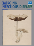
Beatrix Potter, Author, Naturalist, Mycologist [PDF - 711 KB - 2 pages]
| EID | Breedlove B. Beatrix Potter, Author, Naturalist, Mycologist. Emerg Infect Dis. 2019;25(9):1786-1787. https://doi.org/10.3201/eid2509.ac2509 |
|---|---|
| AMA | Breedlove B. Beatrix Potter, Author, Naturalist, Mycologist. Emerging Infectious Diseases. 2019;25(9):1786-1787. doi:10.3201/eid2509.ac2509. |
| APA | Breedlove, B. (2019). Beatrix Potter, Author, Naturalist, Mycologist. Emerging Infectious Diseases, 25(9), 1786-1787. https://doi.org/10.3201/eid2509.ac2509. |


