Volume 12, Number 2—February 2006
Dispatch
Evaluation of a Direct, Rapid Immunohistochemical Test for Rabies Diagnosis
Figure 1
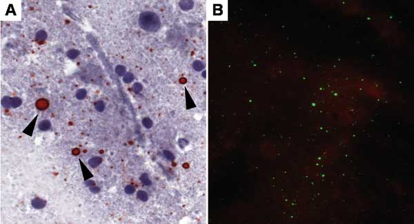
Figure 1. Touch impression of a rabies-positive Tanzanian domestic dog brain preserved in 50% glycerol saline solution for 15 months before testing by direct rapid immunohistochemical test (dRIT) and retested by direct fluorescent-antibody assay (DFA) after 5 months. A) Brain stained by dRIT: rabies virus antigen appears as magenta inclusions (arrowheads) against the blue neuronal hematoxylin counterstain. Magnification, ×630. B) Immunofluorescent apple-green viral inclusions in the same brain processed by DFA. Magnification, ×200.
Page created: February 02, 2012
Page updated: February 02, 2012
Page reviewed: February 02, 2012
The conclusions, findings, and opinions expressed by authors contributing to this journal do not necessarily reflect the official position of the U.S. Department of Health and Human Services, the Public Health Service, the Centers for Disease Control and Prevention, or the authors' affiliated institutions. Use of trade names is for identification only and does not imply endorsement by any of the groups named above.