Skip directly to site content Skip directly to page options Skip directly to A-Z link Skip directly to A-Z link Skip directly to A-Z link
Volume 23, Number 3—March 2017
Research

Zika Virus RNA Replication and Persistence in Brain and Placental Tissue

Julu BhatnagarComments to Author , Demi B. Rabeneck, Roosecelis B. Martines, Sarah Reagan-Steiner, Yokabed Ermias, Lindsey B.C. Estetter, Tadaki Suzuki, Jana M. Ritter, M. Kelly Keating, Gillian Hale, Joy Gary, Atis Muehlenbachs, Amy J. Lambert, Robert Lanciotti, Titilope Oduyebo, Dana Meaney-Delman, Fernando Bolaños, Edgar Alberto Parra Saad, Wun-Ju Shieh, and Sherif Zaki
Author affiliations: Centers for Disease Control and Prevention, Atlanta, Georgia, USA (J. Bhatnagar, D.B. Rabeneck, R.B. Martines, S. Reagan-Steiner, Y. Ermias, L.B.C. Estetter, T. Suzuki, J. Ritter, M.K. Keating, G. Hale, J. Gary, A. Muehlenbachs, T. Oduyebo, D. Meaney-Delman, W. Shieh, S.R. Zaki); Centers for Disease Control and Prevention, Fort Collins, Colorado, USA (A. Lambert, R. Lanciotti); Patología Hospital Universitario de Neiva, Neiva, Colombia (F. Bolaños); Instituto Nacional de Salud, Bogota, Colombia (E.A. Parra Saad)

Main Article

Figure 2

Localization of Zika virus RNA by in situ hybridization in brain tissues from infants with microcephaly. A) ISH with use of antisense probe. Zika virus genomic RNA (red stain) in cerebral cortex of an infant (case-patient no. 66, gestational age 26 wk). Original magnification ×10. B) ISH with use of sense probe. Serial section showing negative-strand replicative RNA intermediates (red stain) in the same areas shown in panel A. Original magnification ×10. C) ISH with use of antisense probe. Highe

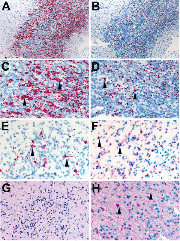
Figure 2. Localization of Zika virus RNA by in situ hybridization in brain tissues from infants with microcephaly. A) ISH with use of antisense probe. Zika virus genomic RNA (red stain) in cerebral cortex of an infant (case-patient no. 66, gestational age 26 wk). Original magnification ×10. B) ISH with use of sense probe. Serial section showing negative-strand replicative RNA intermediates (red stain) in the same areas shown in panel A. Original magnification ×10. C) ISH with use of antisense probe. Higher magnification of panel A, showing cytoplasmic staining of neural (arrowheads) and glial cells. Original magnification ×20. D) ISH with use of sense probe. Higher magnification of panel B, showing cytoplasmic staining of neural and glial cells (arrowheads). Original magnification ×20. E) ISH with use of antisense probe. Localization of negative-strand replicative RNA intermediates in neural cells or neurons (red, arrowheads) of another infant with fatal outcome (case-patient no. 67, gestational age 27 wk). Original magnification ×40. F) Immunostaining of neurons (arrowheads) with use of antibodies against neuronal nuclei in a serial section. Original magnification ×40. G) Hematoxylin and eosin stain showing cortical neural cells in a serial section. Original magnification ×40. H) Immunostaining of glial cells (arrowheads) with use of glial fibrillary acidic protein antibody in the same case. Original magnification ×40. ISH, in situ hybridization.

Main Article

Page created: February 17, 2017
Page updated: February 17, 2017
Page reviewed: February 17, 2017
The conclusions, findings, and opinions expressed by authors contributing to this journal do not necessarily reflect the official position of the U.S. Department of Health and Human Services, the Public Health Service, the Centers for Disease Control and Prevention, or the authors' affiliated institutions. Use of trade names is for identification only and does not imply endorsement by any of the groups named above.
file_external