Skip directly to site content Skip directly to page options Skip directly to A-Z link Skip directly to A-Z link Skip directly to A-Z link
Volume 27, Number 1—January 2021
Research

Cellular Immunity in COVID-19 Convalescents with PCR-Confirmed Infection but with Undetectable SARS-CoV-2–Specific IgG

Sina Schwarzkopf, Adalbert Krawczyk, Dietmar Knop, Hannes Klump, Andreas Heinold, Falko M. Heinemann, Laura Thümmler, Christian Temme, Marianne Breyer, Oliver Witzke, Ulf Dittmer, Veronika Lenz, Peter A. Horn, and Monika LindemannComments to Author 
Author affiliations: Institute for Transfusion Medicine, University Hospital Essen, University of Duisburg-Essen, Essen, Germany (S. Schwarzkopf, D. Knop, H. Klump, A. Heinold, F.M. Heinemann, L. Thümmler, C. Temme, M. Breyer, V. Lenz, P.A. Horn, M. Lindemann); Department of Infectious Diseases, West German Centre of Infectious Diseases, Universitätsmedizin Essen, University of Duisburg-Essen, Essen (A. Krawczyk, O. Witzke); Institute for Virology, University Hospital Essen, University of Duisburg-Essen, Essen (U. Dittmer)

Main Article

Figure 2

Cellular immunity against severe acute respiratory syndrome coronavirus 2 (SARS-CoV-2) as determined by ELISpot assay in potential convalescent-plasma donors with PCR-confirmed infection, Germany. Peripheral blood mononuclear cells of volunteers were stimulated by an S1 protein antigen of SARS-CoV-2 (A) and by peptide pools of the S1/S2 (B) and the M protein (C). If volunteers were tested sequentially, we only included the first dataset. The 3 left groups represent potential convalescent-plasma donors with PCR-confirmed SARS-CoV-2 infection. They either had a strong positive antibody response to the SARS-CoV-2 IgG ELISA as defined by an antibody ratio (R) of >3 (n = 15), an intermediate response (ratio of 1.1–3, n = 4) or borderline or negative results (ratio of <1.1, n = 9). The right group displays data in healthy controls without symptoms of respiratory or gastrointestinal infections and without household contact with SARS-CoV-2 infected patients since January 2020 (n = 22). The group has tested negative or has not been tested by SARS-CoV-2 PCR. Responses in the 4 groups of volunteers were compared by Kruskal-Wallis test with Dunn’s correction. Dotted lines represent 3 spots increment. Horizontal lines indicate median values. Stimulation by S1 protein could not be performed in 7 volunteers; stimulation by the M peptide pool could not be performed in 6. Red dots represent volunteers with an antibody ratio >3; black dots, volunteers with a ratio of 1.1–3; blue dots, volunteers with ratio <1.1; green, NC. IFN-γ, interferon-γ; NC, negative controls.

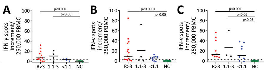
Figure 2. Cellular immunity against severe acute respiratory syndrome coronavirus 2 (SARS-CoV-2) as determined by ELISpot assay in potential convalescent-plasma donors with PCR-confirmed infection, Germany. Peripheral blood mononuclear cells of volunteers were stimulated by an S1 protein antigen of SARS-CoV-2 (A) and by peptide pools of the S1/S2 (B) and the M protein (C). If volunteers were tested sequentially, we only included the first dataset. The 3 left groups represent potential convalescent-plasma donors with PCR-confirmed SARS-CoV-2 infection. They either had a strong positive antibody response to the SARS-CoV-2 IgG ELISA as defined by an antibody ratio (R) of >3 (n = 15), an intermediate response (ratio of 1.1–3, n = 4) or borderline or negative results (ratio of <1.1, n = 9). The right group displays data in healthy controls without symptoms of respiratory or gastrointestinal infections and without household contact with SARS-CoV-2 infected patients since January 2020 (n = 22). The group has tested negative or has not been tested by SARS-CoV-2 PCR. Responses in the 4 groups of volunteers were compared by Kruskal-Wallis test with Dunn’s correction. Dotted lines represent 3 spots increment. Horizontal lines indicate median values. Stimulation by S1 protein could not be performed in 7 volunteers; stimulation by the M peptide pool could not be performed in 6. Red dots represent volunteers with an antibody ratio >3; black dots, volunteers with a ratio of 1.1–3; blue dots, volunteers with ratio <1.1; green, NC. IFN-γ, interferon-γ; NC, negative controls.

Main Article

Page created: October 15, 2020
Page updated: December 21, 2020
Page reviewed: December 21, 2020
The conclusions, findings, and opinions expressed by authors contributing to this journal do not necessarily reflect the official position of the U.S. Department of Health and Human Services, the Public Health Service, the Centers for Disease Control and Prevention, or the authors' affiliated institutions. Use of trade names is for identification only and does not imply endorsement by any of the groups named above.
file_external