Successful Treatment of Balamuthia mandrillaris Granulomatous Amebic Encephalitis with Nitroxoline
Natasha Spottiswoode

, Douglas Pet, Annie Kim, Katherine Gruenberg, Maulik Shah, Amrutha Ramachandran, Matthew T. Laurie, Maham Zia, Camille Fouassier, Christine L. Boutros, Rufei Lu, Yueyuan Zhang, Venice Servellita, Andrew Bollen, Charles Y. Chiu, Michael R. Wilson, Liza Valdivia
1, and Joseph L. DeRisi
1
Author affiliations: University of California, San Francisco, California, USA (N. Spottiswoode, D. Pet, A. Kim, K. Gruenberg, M. Shah, Amrutha Ramachandran, Matthew T. Laurie, Maham Zia, Camille Fouassier, Christine L. Boutros, Rufei Lu, Yueyuan Zhang, Venice Servellita, Andrew Bollen, Charles Y. Chiu, Michael R. Wilson, Liza Valdivia, Joseph L. DeRisi); Chan Zuckerberg Biohub, San Francisco (Joseph L. DeRisi)
Main Article
Figure 2
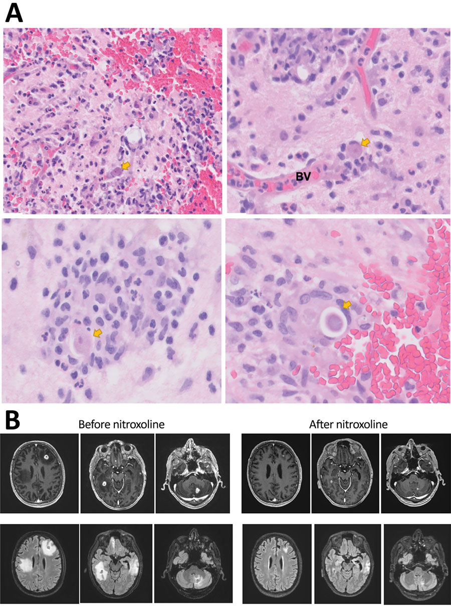
Figure 2. Diagnostic findings for patient with granulomatous amebic encephalitis, California, USA. A) Brain biopsy sample. Granulomas are noted in a perivascular pattern. Scattered structures (arrows) with large nuclei and abundant cytoplasma are concerning for amebic trophozoite forms. Occasional structures with a large nucleus present within a relatively rigid outline (lower right image) are suspicious for amebic cysts, the dormant, thick-walled life stage. B) Magnetic resonance images obtained before and after nitroxoline treatment. Upper row shows axial gadolinium-enhanced T1-weighted images; lower row shows axial fluid-attenuated inversion recovery images. Images in the left series were obtained on day 96 after initial visit, 1 week before nitroxoline initiation; images in the right series were obtained on day 156 after initial visit, 7 weeks after nitroxoline initiation. BV, blood vessel.
Main Article
Page created: November 30, 2022
Page updated: December 22, 2022
Page reviewed: December 22, 2022
The conclusions, findings, and opinions expressed by authors contributing to this journal do not necessarily reflect the official position of the U.S. Department of Health and Human Services, the Public Health Service, the Centers for Disease Control and Prevention, or the authors' affiliated institutions. Use of trade names is for identification only and does not imply endorsement by any of the groups named above.