Volume 29, Number 12—December 2023
Research
Fatal Human Neurologic Infection Caused by Pigeon Avian Paramyxovirus-1, Australia
Siobhan Hurley1 , John Sebastian Eden1, John Bingham, Michael Rodriguez, Matthew J. Neave, Alexandra Johnson, Annaleise R. Howard-Jones, Jen Kok, Antoinette Anazodo, Brendan McMullan, David T. Williams, James Watson, Annalisa Solinas, Ki Wook Kim2, and William Rawlinson2
Figure 1
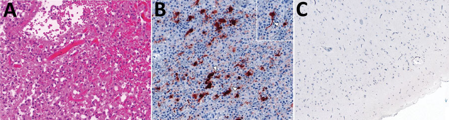
Figure 1. Histology of brain tissue and imaging from a fatal neurologic infection in an immunocompromised child in Australia that was caused by APMV-1. A) Brain biopsy showing extensive cortical necrosis with a dense infiltrate of macrophages. Hematoxylin and eosin stain; original magnification ×20. B) Immunohistochemistry of brain biopsy showing cytoplasmic APMV-1 nucleoprotein, probably in neurons, with axon-like processes (arrowheads). Original magnification ×20 (inset ×40). C) Immunohistochemistry of APMV-1 nucleoprotein, demonstrating the absence of immunolabeling in normal brain tissue. APMV, avian paramyxovirus.
1These first authors contributed equally to this article.
2These senior authors contributed equally to this article.
Page created: November 13, 2023
Page updated: November 18, 2023
Page reviewed: November 18, 2023
The conclusions, findings, and opinions expressed by authors contributing to this journal do not necessarily reflect the official position of the U.S. Department of Health and Human Services, the Public Health Service, the Centers for Disease Control and Prevention, or the authors' affiliated institutions. Use of trade names is for identification only and does not imply endorsement by any of the groups named above.